
Shopping Cart
Remove All Your shopping cart is currently empty

Synonyms: heat shock 70kDa protein 1A
Anti-HSP70 Polyclonal Antibody
| Pack Size | Price | USA Stock | Global Stock | Quantity |
|---|---|---|---|---|
| 50 µL | $220 | 7-10 days | 7-10 days | |
| 100 µL | $374 | 7-10 days | 7-10 days | |
| 200 µL | $527 | 7-10 days | 7-10 days |
| Description | Anti-HSP70 Polyclonal Antibody is a Rabbit antibody targeting HSP70. Anti-HSP70 Polyclonal Antibody can be used in FCM, ICC/IF, IF, IHC-Fr, IHC-P, WB. |
| Synonyms | heat shock 70kDa protein 1A |
| Ig Type | IgG |
| Reactivity | Human,Mouse,Rat (predicted:Chicken,Cow,Rabbit,Sheep) |
| Verified Activity | 1. Blank control (blue line): Jurkat (blue). Primary Antibody (green line): Rabbit Anti-HSP70 antibody (TMAB-00893) Dilution: 1 μg/10^6 cells; Isotype Control Antibody (orange line): Rabbit IgG. Secondary Antibody (white blue line): Goat anti-rabbit IgG-FITC Dilution: 1 μg/test. Protocol The cells were fixed with 2% paraformaldehyde (10 min), then permeabilized with 90% ice-cold methanol for 30 min on ice. Cells stained with Primary Antibody for 30 min at room temperature. The cells were then incubated in 1 X PBS/2% BSA/10% goat serum to block non-specific protein-protein interactions followed by the antibody for 15 min at room temperature. The secondary antibody used for 40 min at room temperature. 2. Hela cell; 4% Paraformaldehyde-fixed; Triton X-100 at room temperature for 20 min; Blocking buffer (normal goat serum) at 37°C for 20 min; Antibody incubation with (HSP70) polyclonal Antibody, Unconjugated (TMAB-00893) 1:100, 90 minutes at 37°C; followed by a conjugated Goat Anti-Rabbit IgG antibody at 37°C for 90 minutes, DAPI (blue) was used to stain the cell nucleus. 3. Tissue/cell: A549 cell; 4% Paraformaldehyde-fixed; Triton X-100 at room temperature for 20 min; Blocking buffer (normal goat serum) at 37°C for 20 min; Antibody incubation with (HSP70) polyclonal Antibody, Unconjugated (TMAB-00893) 1:100, 90 minutes at 37°C; followed by a FITC conjugated Goat Anti-Rabbit IgG antibody at 37°C for 90 minutes, DAPI (blue) was used to stain the cell nucleus. 4. Sample: Lane 1: Testis (Mouse) Lysate at 40 μg Lane 2: Epididymis (Mouse) Lysate at 40 μg Lane 3: Seminal vesicle (Mouse) Lysate at 40 μg Lane 4: Liver (Mouse) Lysate at 40 μg Lane 5: NIH/3T3 (Mouse) Cell Lysate at 30 μg Lane 6: Testis (Rat) Lysate at 40 μg Lane 7: Spleen (Rat) Lysate at 40 μg Lane 8: Hela (Human) Cell Lysate at 30 μg Lane 9: Jurkat (Human) Cell Lysate at 30 μg Lane 10: HepG2 (Human) Cell Lysate at 30 μg Lane 11: A549 (Human) Cell Lysate at 30 μg Lane 12: MCF-7 (Human) Cell Lysate at 30 μg Primary: Anti-HSP70 (TMAB-00893) at 1/1000 dilution Secondary: IRDye800CW Goat Anti-Rabbit IgG at 1/20000 dilution Predicted band size: 70 kDa Observed band size: 68 kDa 5. 25 μg total protein per Lane of various lysates probed with HSP70 polyclonal antibody, unconjugated (TMAB-00893) at 1:1000 dilution and 4°C overnight incubation. Followed by conjugated secondary antibody incubation at RT for 60 min. 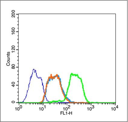 , ,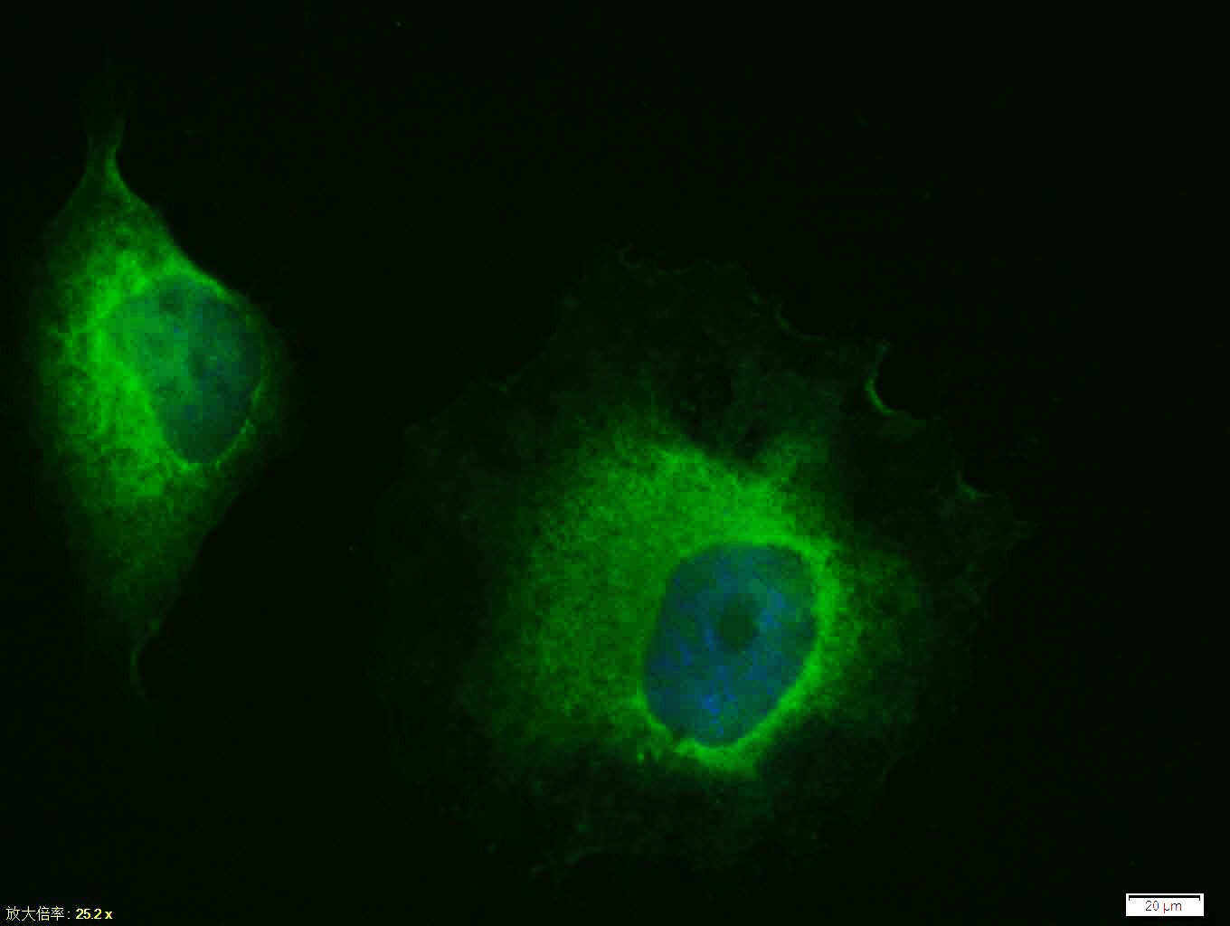 , ,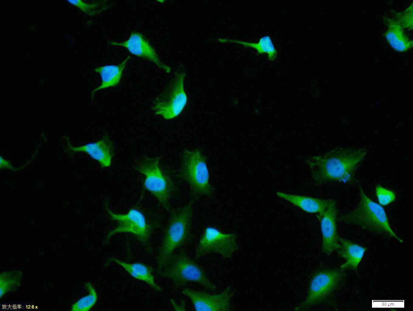 , ,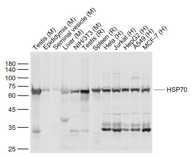 , ,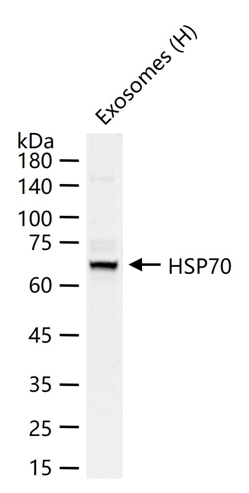 |
| Application | |
| Recommended Dose | FCM=1 μg/Test; ICC/IF=1:100-500; IF=1:100-500; IHC-Fr=1:100-500; IHC-P=1:100-500; WB=1:500-2000 |
| Antibody Type | Polyclonal |
| Host Species | Rabbit |
| Subcellular Localization | Cytoplasm. Note=Localized in cytoplasmic mRNP granules containing untranslated mRNAs. |
| Tissue Specificity | HSPA1B is testis-specific. |
| Construction | Polyclonal Antibody |
| Purification | Protein A purified |
| Appearance | Liquid |
| Formulation | 0.01M TBS (pH7.4) with 1% BSA, 0.02% Proclin300 and 50% Glycerol. |
| Concentration | 1 mg/mL |
| Research Background | This intronless gene encodes a 70kDa heat shock protein which is a member of the heat shock protein 70 family. In conjuction with other heat shock proteins, this protein stabilizes existing proteins against aggregation and mediates the folding of newly translated proteins in the cytosol and in organelles. It is also involved in the ubiquitin-proteasome pathway through interaction with the AU-rich element RNA-binding protein 1. The gene is located in the major histocompatibility complex class III region, in a cluster with two closely related genes which encode similar proteins. [provided by RefSeq, Jul 2008]. |
| Immunogen | KLH conjugated synthetic peptide: human HSP70 |
| Antigen Species | Human |
| Gene Name | HSPA1A |
| Gene ID | |
| Protein Name | Heat shock 70 kDa protein 1A |
| Uniprot ID | |
| Biology Area | Heat Shock Proteins,Tumor biomarkers |
| Function | In cooperation with other chaperones, Hsp70s stabilize preexistent proteins against aggregation and mediate the folding of newly translated polypeptides in the cytosol as well as within organelles. These chaperones participate in all these processes through their ability to recognize nonnative conformations of other proteins. They bind extended peptide segments with a net hydrophobic character exposed by polypeptides during translation and membrane translocation, or following stress-induced damage. In case of rotavirus A infection, serves as a post-attachment receptor for the virus to facilitate entry into the cell. |
| Molecular Weight | Theoretical: 70 kDa. Actual: 68 kDa. |
| Stability & Storage | Store at -20°C or -80°C for 12 months. Avoid repeated freeze-thaw cycles. |
| Transport | Shipping with blue ice. |
| Size | Quantity | Unit Price | Amount | Operation |
|---|
 Hello! How can I help you today?
 Copyright © 2015-2026 TargetMol Chemicals Inc. All Rights Reserved.
