
Shopping Cart
Remove All Your shopping cart is currently empty

Synonyms: WTS, RPD3, MRXS6, histone deacetylase 8, HDACL1, HD8, CDLS5, CDA07
Anti-HDAC8 Antibody
(6A120)
| Pack Size | Price | USA Stock | Global Stock | Quantity |
|---|---|---|---|---|
| 50 µL | $296 | 7-10 days | 7-10 days | |
| 100 µL | $497 | 7-10 days | 7-10 days |
| Description | Anti-HDAC8 Antibody (6A120) is a Rabbit antibody targeting HDAC8. Anti-HDAC8 Antibody (6A120) can be used in FCM,ICC/IF,IHC,IP,WB. |
| Synonyms | WTS, RPD3, MRXS6, histone deacetylase 8, HDACL1, HD8, CDLS5, CDA07 |
| Ig Type | IgG |
| Clone | 6A120 |
| Reactivity | Human |
| Verified Activity | 1. Western blot analysis of HDAC8 on different lysates using anti-HDAC8 antibody at 1/1,000 dilution. Positive control: Lane 1: Hela, Lane 2: K562. 2. Immunohistochemical analysis of paraffin-embedded human lung cancer tissue using anti-HDAC8 antibody. Counter stained with hematoxylin. 3. Immunohistochemical analysis of paraffin-embedded human kidney tissue using anti-HDAC8 antibody. Counter stained with hematoxylin. 4. Immunohistochemical analysis of paraffin-embedded human pancreas tissue using anti-HDAC8 antibody. Counter stained with hematoxylin. 5. Immunohistochemical analysis of paraffin-embedded human lung tissue using anti-HDAC8 antibody. Counter stained with hematoxylin. 6. ICC staining HDAC8 in Hela cells (green). The nuclear counter stain is DAPI (blue). Cells were fixed in paraformaldehyde, permeabilised with 0.25% Triton X100/PBS. 7. ICC staining HDAC8 in A549 cells (green). The nuclear counter stain is DAPI (blue). Cells were fixed in paraformaldehyde, permeabilised with 0.25% Triton X100/PBS. 8. ICC staining HDAC8 in HepG2 cells (green). The nuclear counter stain is DAPI (blue). Cells were fixed in paraformaldehyde, permeabilised with 0.25% Triton X100/PBS. 9. Flow cytometric analysis of K562 cells with HDAC8 antibody at 1/50 dilution (red) compared with an unlabelled control (cells without incubation with primary antibody; black). Alexa Fluor 488-conjugated goat anti rabbit IgG was used as the secondary antibody. 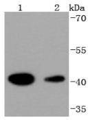 , ,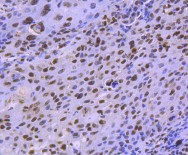 , ,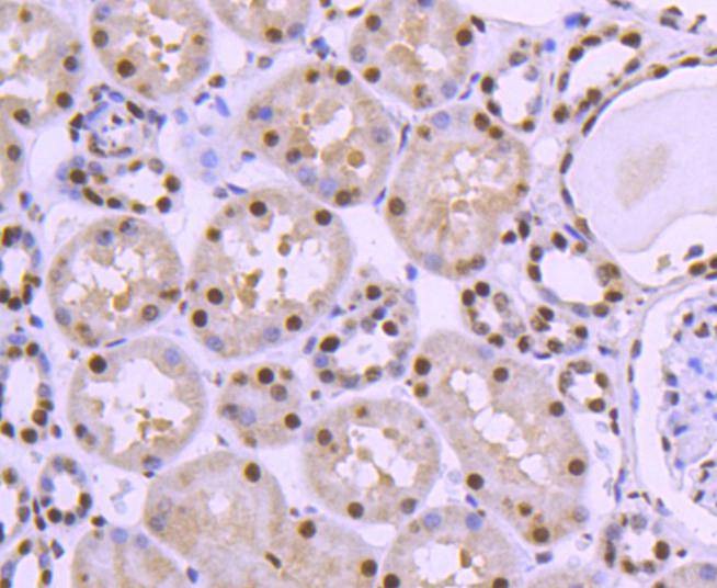 , ,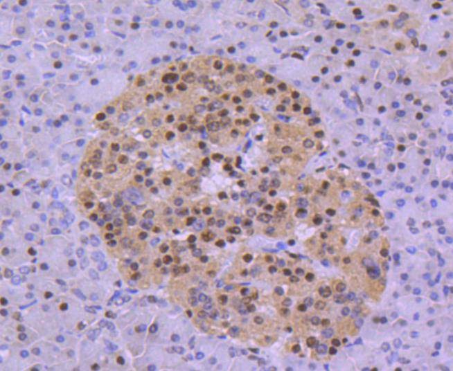 , ,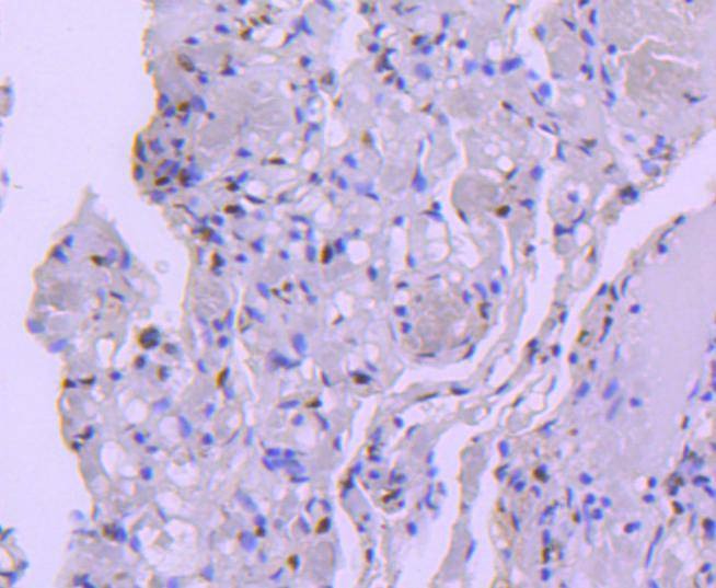 , ,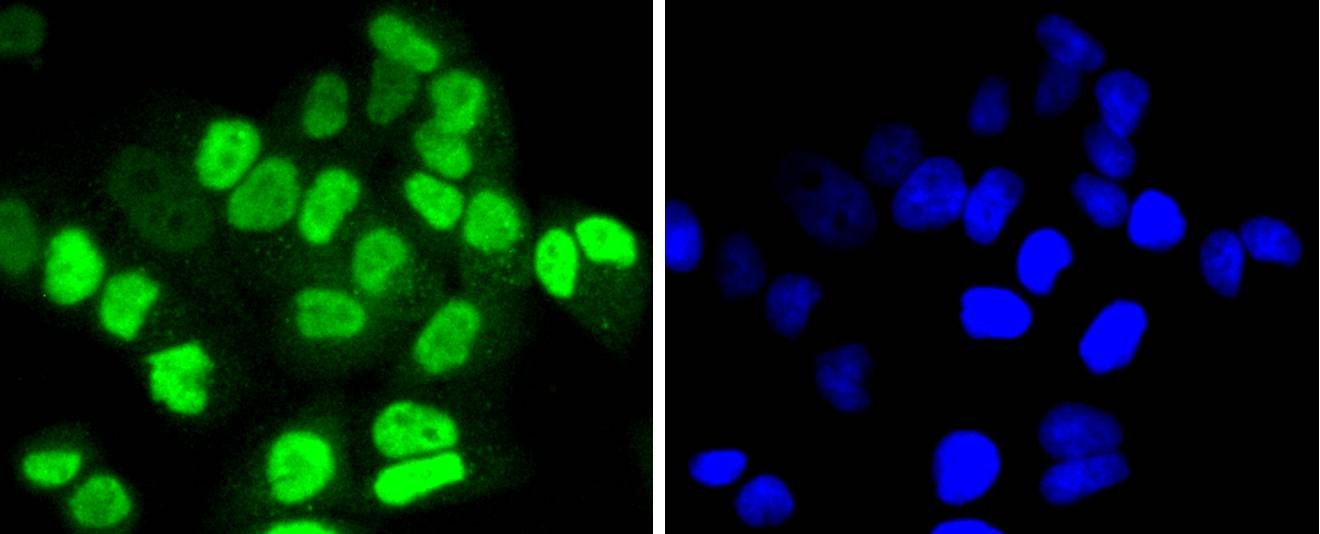 , ,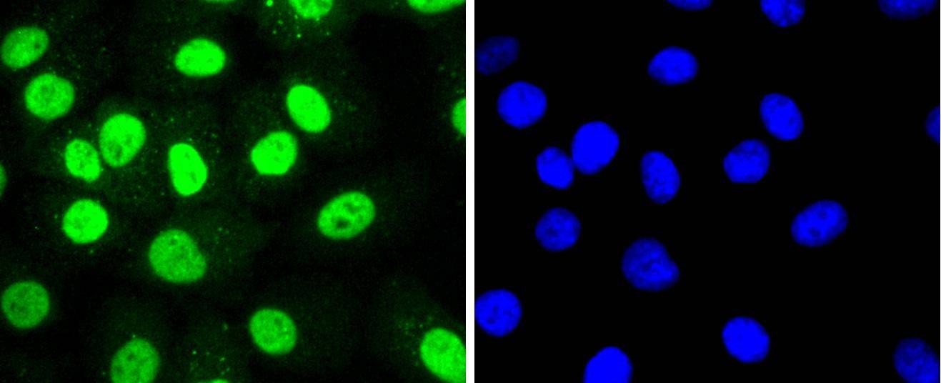 , ,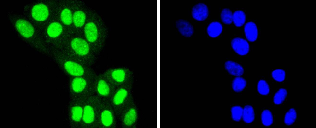 , ,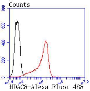 |
| Application | |
| Recommended Dose | WB: 1:1000-2000; IHC: 1:50-200; ICC/IF: 1:100-500; FCM: 1:50-100 |
| Antibody Type | Monoclonal |
| Host Species | Rabbit |
| Construction | Recombinant Antibody |
| Purification | ProA affinity purified |
| Appearance | Liquid |
| Formulation | 1*TBS (pH7.4), 1%BSA, 40%Glycerol. Preservative: 0.05% Sodium Azide. |
| Research Background | In the intact cell, DNA closely associates with histones and other nuclear proteins to form chromatin. The remodeling of chromatin is believed to be a critical component of transcriptional regulation and a major source of this remodeling is brought about by the acetylation of nucleosomal histones. Acetylation of lysine residues in the amino terminal tail domain of histone results in an allosteric change in the nucleosomal conformation and an increased accessibility to transcription factors by DNA. Conversely, the deacetylation of histones is associated with transcriptional silencing. Several mammalian proteins have been identified as nuclear histone acetylases, including GCN5, PCAF (p300/CBP-associated factor), p300/CBP, HAT1 and the TFIID subunit TAF II p250. Mammalian HDAC8, isolated from human kidney, is a histone deacetylase that shares homology to other HDACs but has different tissue distribution. HDAC8 is localized to the nucleus and plays a role in the development of a broad range of tissues and in the etiology of cancer. |
| Conjucates | Unconjugated |
| Immunogen | Recombinant Protein |
| Uniprot ID |
| Molecular Weight | Theoretical: 42 kDa. |
| Stability & Storage | Store at -20°C or -80°C for 12 months. Avoid repeated freeze-thaw cycles. |
| Transport | Shipping with blue ice. |
| Size | Quantity | Unit Price | Amount | Operation |
|---|
 Hello! How can I help you today?
 Copyright © 2015-2026 TargetMol Chemicals Inc. All Rights Reserved.
