
Shopping Cart
Remove All Your shopping cart is currently empty

Synonyms: GJAL, GJA1, Gap junction alpha-1 protein, Gap junction 43 kDa heart protein, Connexin-43 (Cx43)
Anti-GJA1 Polyclonal Antibody
| Pack Size | Price | USA Stock | Global Stock | Quantity |
|---|---|---|---|---|
| 50 µL | $222 | 7-10 days | 7-10 days | |
| 100 µL | $372 | 7-10 days | 7-10 days | |
| 200 µL | $528 | 7-10 days | 7-10 days |
| Description | Anti-GJA1 Polyclonal Antibody is a Rabbit antibody targeting GJA1. Anti-GJA1 Polyclonal Antibody can be used in FCM, ICC/IF, IF, IHC-Fr, IHC-P, WB. |
| Synonyms | GJAL, GJA1, Gap junction alpha-1 protein, Gap junction 43 kDa heart protein, Connexin-43 (Cx43) |
| Ig Type | IgG |
| Reactivity | Human,Mouse,Rat (predicted:Chicken,Dog,Cow) |
| Verified Activity | 1. Paraformaldehyde-fixed, paraffin embedded (Human stomach cancer); Antigen retrieval by boiling in sodium citrate buffer (pH6.0) for 15 min; Block endogenous peroxidase by 3% hydrogen peroxide for 20 min; Blocking buffer (normal goat serum) at 37°C for 30 min; Antibody incubation with (Connexin 43) Polyclonal Antibody, Unconjugated (TMAB-00772) at 1:200 overnight at 4°C, followed by operating according to SP Kit (Rabbit) instructions and DAB staining. 2. Sample: Mcf-7 Cell Lysate at 40 μg Primary: Anti-Connexin (TMAB-00772) at 1/300 dilution Secondary: IRDye800CW Goat Anti-Rabbit IgG at 1/10000 dilution Predicted band size: 42 kDa Observed band size: 43 kDa 3. Tissue/cell: MCF7 cell; 4% Paraformaldehyde-fixed; Triton X-100 at room temperature for 20 min; Blocking buffer (normal goat serum) at 37°C for 20 min; Antibody incubation with (Connexin 43) polyclonal Antibody, Unconjugated (TMAB-00772) 1:100, 90 minutes at 37°C; followed by a FITC conjugated Goat Anti-Rabbit IgG antibody at 37°C for 90 minutes, DAPI (blue) was used to stain the cell nucleus. 4. Tissue/cell: U-251 cell; 4% Paraformaldehyde-fixed; Triton X-100 at room temperature for 20 min; Blocking buffer (normal goat serum) at 37°C for 20 min; Antibody incubation with (Connexin 43) polyclonal Antibody, Unconjugated (TMAB-00772) 1:100, 90 minutes at 37°C; followed by a FITC conjugated Goat Anti-Rabbit IgG antibody at 37°C for 90 minutes, DAPI (blue) was used to stain the cell nucleus. 5. Sample: Lane 1: Heart (Mouse) Lysate at 40 μg Lane 2: Heart (Rat) Lysate at 40 μg Lane 3: Cerebrum (Mouse) Lysate at 40 μg Lane 4: Cerebrum (Rat) Lysate at 40 μg Primary: Anti-Connexin 43 (TMAB-00772) at 1/1000 dilution Secondary: IRDye800CW Goat Anti-Rabbit IgG at 1/20000 dilution Predicted band size: 42 kDa Observed band size: 45 kDa 6. Blank control (blue line): Hela (blue). Primary Antibody (green line): Rabbit Anti-Connexin 43 antibody (TMAB-00772) Dilution: 1 μg/10^6 cells; Isotype Control Antibody (orange line): Rabbit IgG. Secondary Antibody (white blue line): f (ab/)2 fragment goat anti-rabbit IgG-FITC. Dilution: 1 μg/test. Protocol The cells were fixed with 2% paraformaldehyde (10 min), then permeabilized with 90% ice-cold methanol for 30 min on ice. Cells stained with Primary Antibody for 30 min at room temperature. The cells were then incubated in 1 X PBS/2% BSA/10% goat serum to block non-specific protein-protein interactions followed by the antibody for 15 min at room temperature. The secondary antibody used for 40 min at room temperature. 7. Paraformaldehyde-fixed, paraffin embedded (Mouse brain); Antigen retrieval by boiling in sodium citrate buffer (pH6.0) for 15 min; Block endogenous peroxidase by 3% hydrogen peroxide for 20 min; Blocking buffer (normal goat serum) at 37°C for 30 min; Antibody incubation with (Connexin 43) Polyclonal Antibody, Unconjugated (TMAB-00772) at 1:400 overnight at 4°C, followed by operating according to SP Kit (Rabbit) instructionsand DAB staining. 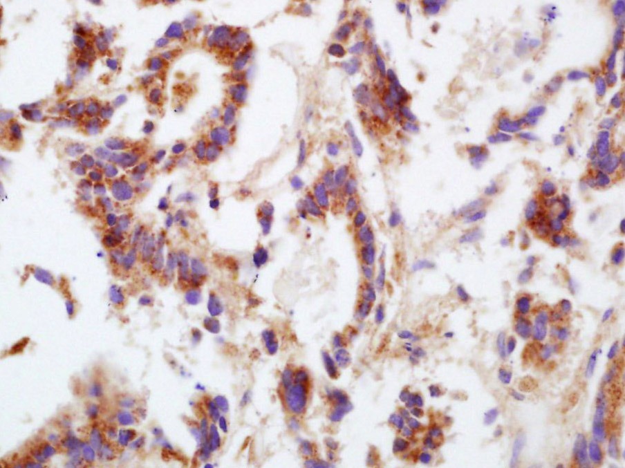 , ,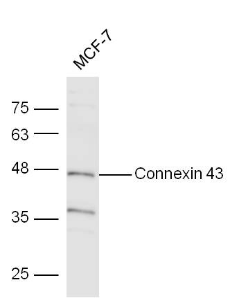 , ,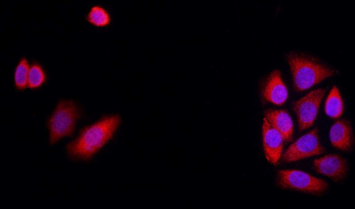 , ,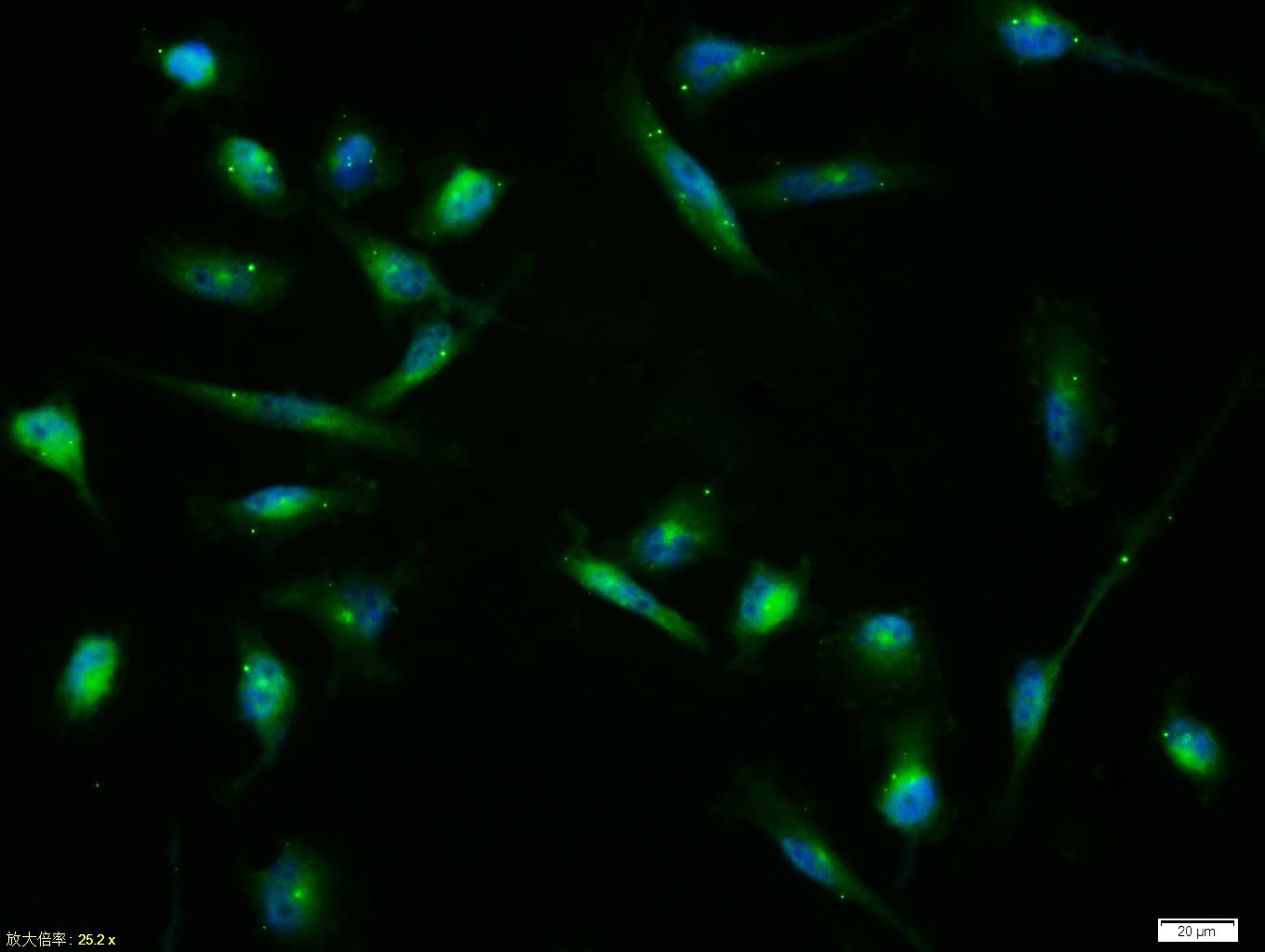 , ,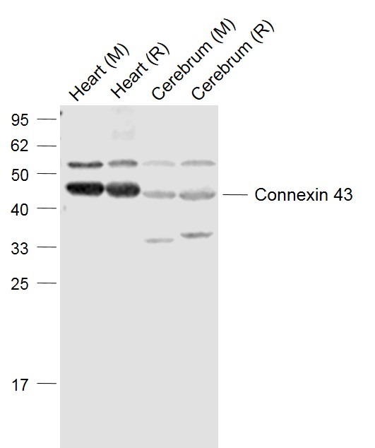 , ,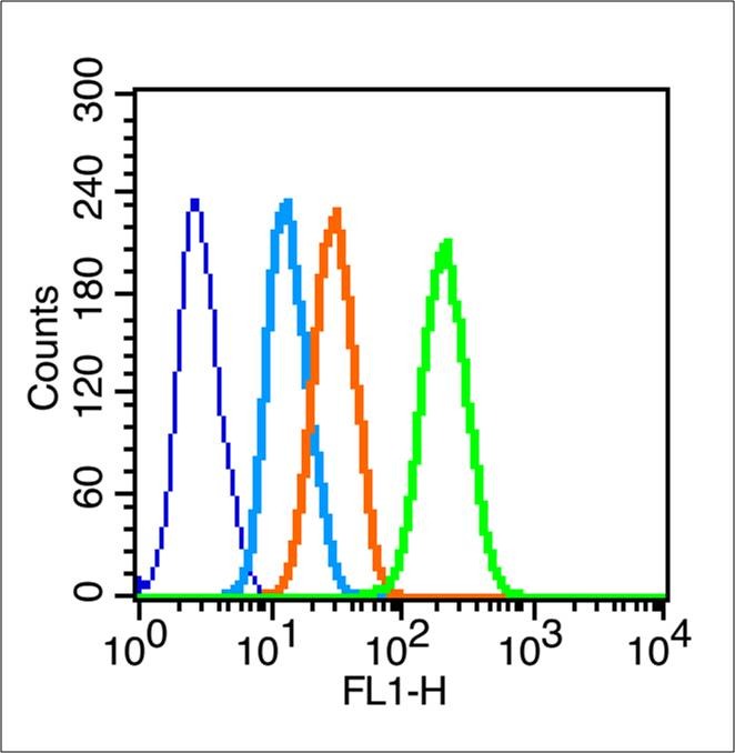 , ,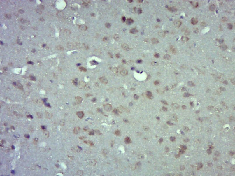 |
| Application | |
| Recommended Dose | FCM=1 μg/Test; ICC/IF=1:100-500; IF=1:100-500; IHC-Fr=1:100-500; IHC-P=1:100-500; WB=1:500-2000 |
| Antibody Type | Polyclonal |
| Host Species | Rabbit |
| Subcellular Localization | Cell membrane; Multi-pass membrane protein. Cell junction, gap junction. |
| Tissue Specificity | Expressed in the heart and fetal cochlea. |
| Construction | Polyclonal Antibody |
| Purification | Protein A purified |
| Appearance | Liquid |
| Formulation | 0.01M TBS (pH7.4) with 1% BSA, 0.02% Proclin300 and 50% Glycerol. |
| Concentration | 1 mg/mL |
| Research Background | This gene is a member of the connexin gene family. The encoded protein is a component of gap junctions, which are composed of arrays of intercellular channels that provide a route for the diffusion of low molecular weight materials from cell to cell. The encoded protein is the major protein of gap junctions in the heart that are thought to have a crucial role in the synchronized contraction of the heart and in embryonic development. A related intronless pseudogene has been mapped to chromosome 5. Mutations in this gene have been associated with oculodentodigital dysplasia and heart malformations. [provided by RefSeq]. |
| Immunogen | KLH conjugated synthetic peptide: human Connexin-43 |
| Antigen Species | Human |
| Gene Name | GJA1 |
| Gene ID | |
| Protein Name | Gap junction alpha-1 protein |
| Uniprot ID | |
| Biology Area | Cell junction molecules,Cardiac arrhythmias,Gap Junctions |
| Function | Gap junction protein that acts as a regulator of bladder capacity. A gap junction consists of a cluster of closely packed pairs of transmembrane channels, the connexons, through which materials of low MW diffuse from one cell to a neighboring cell. May play a critical role in the physiology of hearing by participating in the recycling of potassium to the cochlear endolymph. Negative regulator of bladder functional capacity: acts by enhancing intercellular electrical and chemical transmission, thus sensitizing bladder muscles to cholinergic neural stimuli and causing them to contract. |
| Molecular Weight | Theoretical: 42 kDa. Actual: 45 kDa. |
| Stability & Storage | Store at -20°C or -80°C for 12 months. Avoid repeated freeze-thaw cycles. |
| Transport | Shipping with blue ice. |
| Size | Quantity | Unit Price | Amount | Operation |
|---|
 Hello! How can I help you today?
 Copyright © 2015-2026 TargetMol Chemicals Inc. All Rights Reserved.
