
Shopping Cart
Remove All Your shopping cart is currently empty

Synonyms: GFP Protein
Anti-GFP Antibody
(2A799)
| Pack Size | Price | USA Stock | Global Stock | Quantity |
|---|---|---|---|---|
| 100 µL | $106 | 7-10 days | 7-10 days | |
| 200 µL | $183 | 7-10 days | 7-10 days | |
| 500 µL | $428 | 7-10 days | 7-10 days | |
| 1 mL | $804 | 7-10 days | 7-10 days |
| Description | Anti-GFP Antibody (2A799) is a Mouse antibody targeting Phospho-IRF3 (Ser386). Anti-GFP Antibody (2A799) can be used in WB,ELISA,IHC-P,IHC-F,IF,ICC/IF,FCM,IP. |
| Synonyms | GFP Protein |
| Ig Type | IgG |
| Clone | 2A799 |
| Reactivity | Species independent |
| Verified Activity | 1. Transformed (GFP or EV) E. coli cells lysates were subjected to SDS-PAGE followed by WB with TMAB-00764 (Anti-GFP) at dilution of 1:1,000,000 incubated at 4°C overnight. 2. Sample: GFP-tagged fusion protein Overexpression E.coli Lysate at 4 μg Primary: Anti-GFP-Tag (TMAB-00764) at 1/1000000 dilution Secondary: IRDye800CW Goat Anti-Mouse IgG at 1/20000 dilution Predicted band size: 28 kDa Observed band size: 30 kDa 3. Sample: GFP-Tagged Fusion Protein Overexpression E.coli Lysate at 2 μg Primary: Anti-GST tag (TMAB-00764) at 1/5000 ~ 1/10000 dilution Secondary: IRDye800CW Goat Anti-Mouse IgG at 1/20000 dilution Predicted band size: 28 kDa Observed band size: 28 kDa 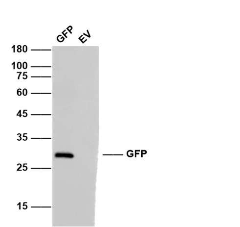 , ,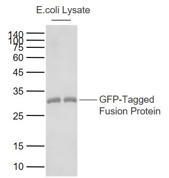 , ,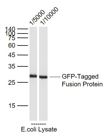 |
| Application | |
| Recommended Dose | WB=1:50000-500000,ELISA=1:1000-5000,IHC-P=1:200-1000,IHC-F=1:200-1000,IF=1:200-1000,ICC/IF=1:100-500,FCM=1ug/Test,IP=1:50-200 |
| Antibody Type | Monoclonal |
| Host Species | Mouse |
| Tissue Specificity | Photocytes. |
| Construction | Hybridoma Monoclonal Antibody |
| Purification | Protein G purified |
| Appearance | Liquid |
| Formulation | 0.01M TBS (pH7.4) with 1% BSA, 0.02% Proclin300 and 50% Glycerol. |
| Concentration | 1 mg/mL |
| Research Background | Green fluorescence protein (GFP) is a 27 kDa protein derived from the jellyfish Aequorea victoria, which emits green light (emission peak at a wavelenth of 509 nm) when excited by blue light (excitation peak at a wavelenth of 395 nm). Green Fluorescent Protein (GFP) has become an invaluable tool in cell biology research, since its intrinsic fluorescence can be visualized in living cells. GFP fluorescence is stable under fixation conditions and suitable for a variety of applications. GFP has been widely used as a reporter for gene expression, enabling researchers to visualize and localize GFP-tagged proteins within living cells without the need for chemical staining. Other applications of GFP include assessment of protein protein interactions through the yeast two hybrid system and measurement of distance between proteins through fluorescence energy transfer (FRET) protocols. GFP technnology has considerably contributed to a greater understanding of cellular physiology. |
| Immunogen | Recombinant Protein: Green Fluorescence Protein |
| Gene Name | GFP |
| Protein Name | Green fluorescent protein |
| Biology Area | GFP |
| Function | Energy-transfer acceptor. Its role is to transduce the blue chemiluminescence of the protein aequorin into green fluorescent light by energy transfer. Fluoresces in vivo upon receiving energy from the Ca(2+)-activated photoprotein aequorin |
| Molecular Weight | Actual: 28-30 kDa. |
| Stability & Storage | Store at -20°C or -80°C for 12 months. Avoid repeated freeze-thaw cycles. |
| Transport | Shipping with blue ice. |
| Size | Quantity | Unit Price | Amount | Operation |
|---|
 Hello! How can I help you today?
 Copyright © 2015-2026 TargetMol Chemicals Inc. All Rights Reserved.
