
Shopping Cart
Remove All Your shopping cart is currently empty

Synonyms: hDH V, FUSE-binding protein 1, FUBP 1, FBP, Far upstream element-binding protein 1, DNA helicase V
Anti-FUBP1 Antibody
(1H129)
| Pack Size | Price | USA Stock | Global Stock | Quantity |
|---|---|---|---|---|
| 50 µL | $298 | 7-10 days | 7-10 days | |
| 100 µL | $498 | 7-10 days | 7-10 days |
| Description | Anti-FUBP1 Antibody (1H129) is a Rabbit antibody targeting FUBP1. Anti-FUBP1 Antibody (1H129) can be used in FCM,ICC/IF,IHC,WB. |
| Synonyms | hDH V, FUSE-binding protein 1, FUBP 1, FBP, Far upstream element-binding protein 1, DNA helicase V |
| Ig Type | IgG |
| Clone | 1H129 |
| Reactivity | Human,Mouse,Rat |
| Verified Activity | 1. Western blot analysis of FUBP1 on different lysates using anti-FUBP1 antibody at 1/1,000 dilution. Positive control: Lane 1: Hela, Lane 2: Raji. 2. Immunohistochemical analysis of paraffin-embedded mouse pancreas tissue using anti-FUBP1 antibody. Counter stained with hematoxylin. 3. Immunohistochemical analysis of paraffin-embedded human breast carcinoma tissue using anti-FUBP1 antibody. Counter stained with hematoxylin. 4. Immunohistochemical analysis of paraffin-embedded human pancreas tissue using anti-FUBP1 antibody. Counter stained with hematoxylin. 5. Immunohistochemical analysis of paraffin-embedded mouse brain tissue using anti-FUBP1 antibody. Counter stained with hematoxylin. 6. Immunohistochemical analysis of paraffin-embedded human tonsil tissue using anti-FUBP1 antibody. Counter stained with hematoxylin. 7. Immunohistochemical analysis of paraffin-embedded mouse stomach tissue using anti-FUBP1 antibody. Counter stained with hematoxylin. 8. ICC staining FUBP1 in Hela cells (green). The nuclear counter stain is DAPI (blue). Cells were fixed in paraformaldehyde, permeabilised with 0.25% Triton X100/PBS. 9. ICC staining FUBP1 in MCF-7 cells (green). The nuclear counter stain is DAPI (blue). Cells were fixed in paraformaldehyde, permeabilised with 0.25% Triton X100/PBS. 10. ICC staining FUBP1 in HepG2 cells (green). The nuclear counter stain is DAPI (blue). Cells were fixed in paraformaldehyde, permeabilised with 0.25% Triton X100/PBS. 11. Flow cytometric analysis of Jurakt cells with FUBP1 antibody at 1/50 dilution (red) compared with an unlabelled control (cells without incubation with primary antibody; black). Alexa Fluor 488-conjugated goat anti rabbit IgG was used as the secondary antibody. 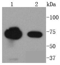 , ,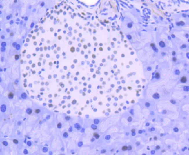 , ,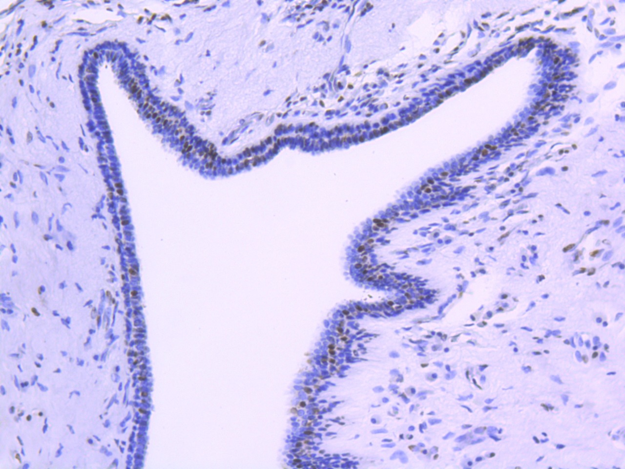 , ,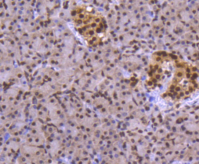 , ,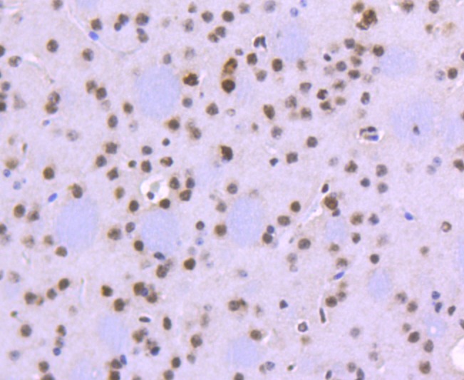 , ,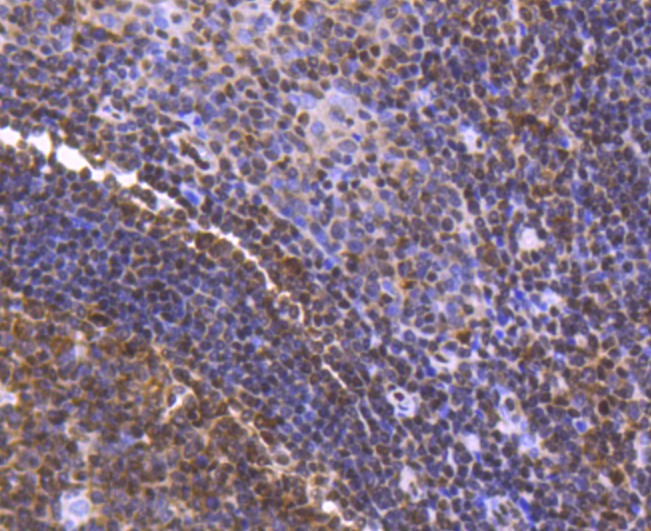 , ,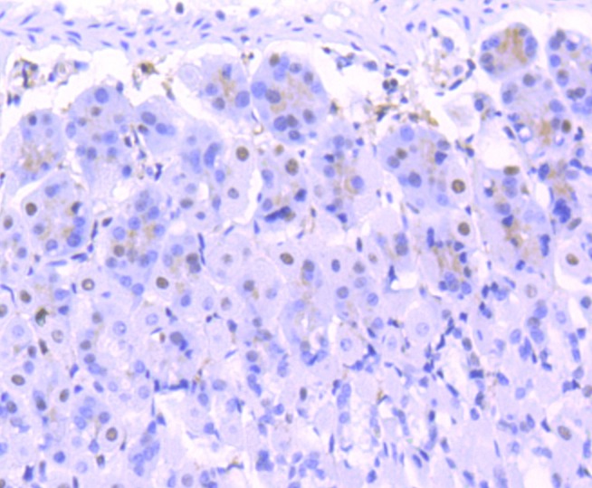 , ,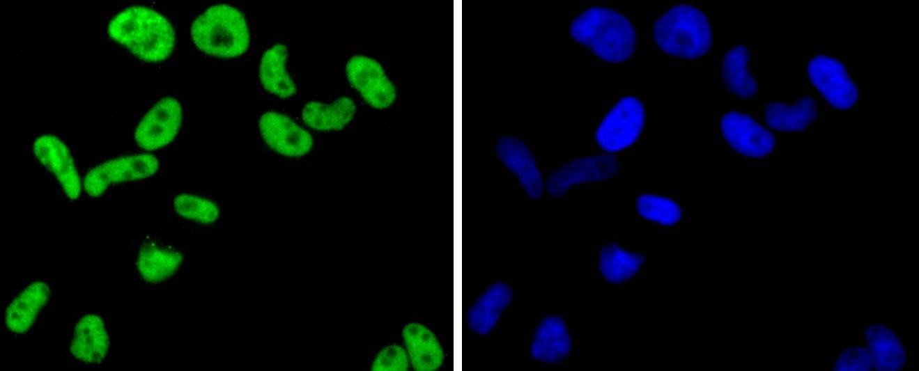 , ,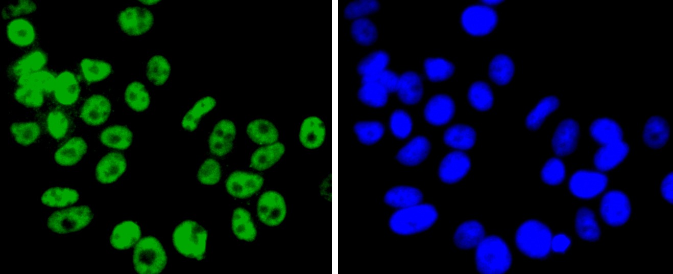 , ,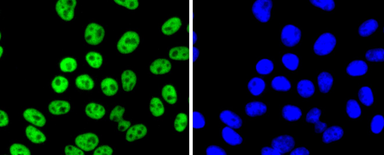 , ,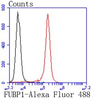 |
| Application | |
| Recommended Dose | WB: 1:1000-2000; IHC: 1:50-200; ICC/IF: 1:100-500; FCM: 1:50-100 |
| Antibody Type | Monoclonal |
| Host Species | Rabbit |
| Construction | Recombinant Antibody |
| Purification | ProA affinity purified |
| Appearance | Liquid |
| Formulation | 1*TBS (pH7.4), 1%BSA, 40%Glycerol. Preservative: 0.05% Sodium Azide. |
| Research Background | Activation of FUSE, the far upstream element, is required for the proper ex-pression of the mammalian gene c-Myc in undifferentiated cells. The binding of FBP1 (FUSE-binding protein or far upstream element-binding protein) to FUSE is necessary for c-Myc expression, indicating that FBP1 functions as a growth-dependent regulator of c-Myc expression. Isolated from proliferating HL-60 cells, FBP1 (FBP), FBP2 and FBP3 comprise a family of single-stranded DNA-binding proteins that specifically bind to FUSE elements. The FBP transcription factors share a conserved central DNA-binding domain and show significant homology in their carboxyl-terminal activation domains. Expression of FBP1 is detected in undifferentiated cells and is substantially decreased following cellular differentiation. |
| Conjucates | Unconjugated |
| Immunogen | Recombinant Protein |
| Uniprot ID |
| Molecular Weight | Theoretical: 74 kDa. |
| Stability & Storage | Store at -20°C or -80°C for 12 months. Avoid repeated freeze-thaw cycles. |
| Transport | Shipping with blue ice. |
| Size | Quantity | Unit Price | Amount | Operation |
|---|
 Hello! How can I help you today?
 Copyright © 2015-2026 TargetMol Chemicals Inc. All Rights Reserved.
