
Shopping Cart
Remove All Your shopping cart is currently empty

Synonyms: TNLG1A, TNFSF6, FASLG, FASL, Fas ligand, CD95-L, CD95L, CD178, APTL, APT1LG1, ALPS1B
Anti-Fas Ligand Polyclonal Antibody
| Pack Size | Price | USA Stock | Global Stock | Quantity |
|---|---|---|---|---|
| 50 µL | $222 | 7-10 days | 7-10 days | |
| 100 µL | $372 | 7-10 days | 7-10 days | |
| 200 µL | $528 | 7-10 days | 7-10 days |
| Description | Anti-Fas Ligand Polyclonal Antibody is a Rabbit antibody targeting Fas Ligand. Anti-Fas Ligand Polyclonal Antibody can be used in FCM,IF,IHC-Fr,IHC-P,WB. |
| Synonyms | TNLG1A, TNFSF6, FASLG, FASL, Fas ligand, CD95-L, CD95L, CD178, APTL, APT1LG1, ALPS1B |
| Ig Type | IgG |
| Reactivity | Human,Mouse,Rat (predicted:Cow) |
| Verified Activity | 1. Sample: Jurkat (Human) Cell Lysate at 30 μg Primary: Anti-Fas Ligand (TMAB-00655) at 1/1000 dilution Secondary: IRDye800CW Goat Anti-Rabbit IgG at 1/20000 dilution Predicted band size: 31 kDa Observed band size: 40 kDa 2. Tissue/cell: human rectal carcinoma;4% Paraformaldehyde-fixed and paraffin-embedded; Antigen retrieval: citrate buffer (0.01M, pH6.0), Boiling bathing for 15 min; Blocking buffer (normal goat serum) at 37°C for 20 min; Incubation: Anti-FasL Polyclonal Antibody, Unconjugated (TMAB-00655) 1:200, overnight at 4°C; The secondary antibody was Goat Anti-Rabbit IgG, Cy3 conjugated used at 1:200 dilution for 40 minutes at 37°C. DAPI (5 μg/ml,blue) was used to stain the cell nucleus. 3. Blank control: mouse thymouses (blue) Isotype Control Antibody: Rabbit Igg (orange); Secondary Antibody: Goat anti-rabbit IgG-FITC (white blue), Dilution: 1:100 in 1 X PBS containing 0.5% BSA; Primary Antibody Dilution: 1 μL in 100 μL 1X PBS containing 0.5% BSA (green). 4. Blank control: Mouse Kidney (blue). Primary Antibody: Rabbit Anti-phospho-Fas Ligand antibody (TMAB-00655,Green); Dilution: 1 μg in 100 μL 1X PBS containing 0.5% BSA; Isotype Control Antibody: Rabbit Igg (orange),used under the same conditions; Secondary Antibody: Goat anti-rabbit IgG-FITC (white blue), Dilution: 1:200 in 1 X PBS containing 0.5% BSA. Protocol The cells were fixed with 2% paraformaldehyde for 10 min at 37°C. Primary antibody (TMAB-00655, 1 μg/1x10^6 cells) were incubated for 30 min at room temperature, followed by 1 X PBS containing 0.5% BSA + 10% goat serum (15 min) to block non-specific protein-protein interactions. Then the Goat Anti-rabbit IgG/FITC antibody was added into the blocking buffer mentioned above to react with the primary antibody at 1/200 dilution for 40 min on ice. 5. Sample: Bone (mouse) Lysate at 40 μg Primary: Anti-Fas ligand (TMAB-00655) at 1/300 dilution Secondary: IRDye800CW Goat Anti-Rabbit IgG at 1/20000 dilution Predicted band size: 31 kDa Observed band size: 46 kDa 6. Sample: Lane 1: Bone (Mouse) Lysate at 40 μg Lane 2: Bone (Rat) Lysate at 40 μg Primary: Anti-Fas Ligand (TMAB-00655) at 1/1000 dilution Secondary: IRDye800CW Goat Anti-Rabbit IgG at 1/20000 dilution Predicted band size: 40 kDa Observed band size: 40 kDa 7. Paraformaldehyde-fixed, paraffin embedded (mouse placenta); Antigen retrieval by boiling in sodium citrate buffer (pH6.0) for 15 min; Block endogenous peroxidase by 3% hydrogen peroxide for 20 min; Blocking buffer (normal goat serum) at 37°C for 30 min; Antibody incubation with (Fas Ligand) Polyclonal Antibody, Unconjugated (TMAB-00655) at 1:200 overnight at 4°C, followed by operating according to SP Kit (Rabbit) instructionsand DAB staining. 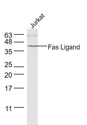 , ,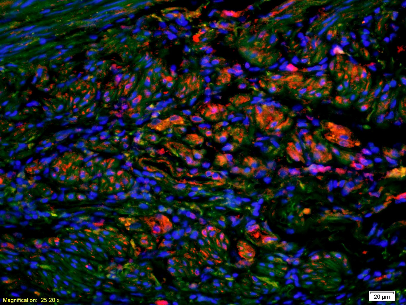 , ,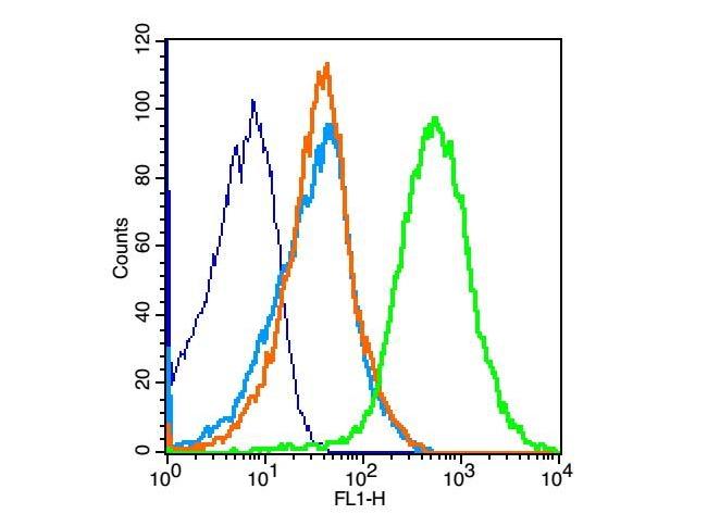 , ,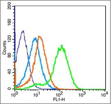 , ,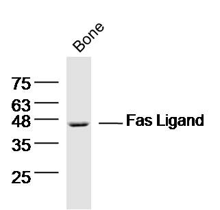 , ,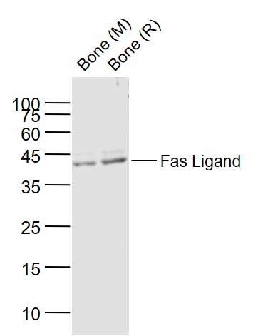 , ,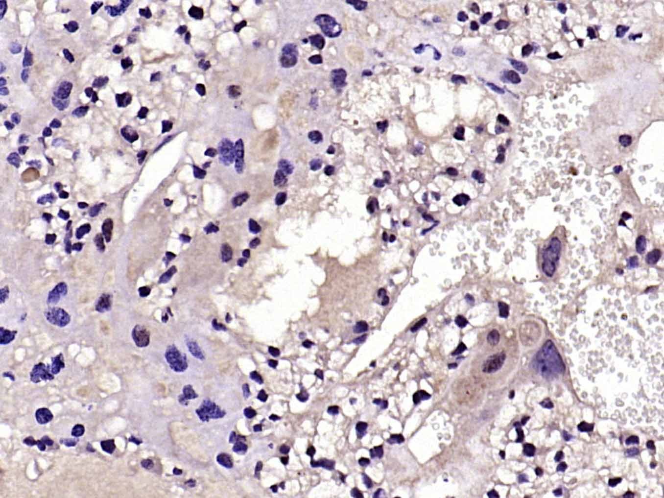 |
| Application | |
| Recommended Dose | WB: 1:500-2000; IHC-P: 1:100-500; IHC-Fr: 1:100-500; IF: 1:100-500; FCM: 1μg/Test |
| Antibody Type | Polyclonal |
| Host Species | Rabbit |
| Subcellular Localization | Cell membrane; Single-pass type II membrane protein. Secreted. Cytoplasmic vesicle lumen. Lysosome lumen. Note=May be released into the extracellular fluid, probably by cleavage form the cell surface. Is internalized into multivesicular bodies of secretory lysosomes after phosphorylation by FGR and monoubiquitination. |
| Construction | Polyclonal Antibody |
| Purification | Protein A purified |
| Appearance | Liquid |
| Formulation | 0.01M TBS (pH7.4) with 1% BSA, 0.02% Proclin300 and 50% Glycerol. |
| Concentration | 1 mg/mL |
| Research Background | This gene is a member of the tumor necrosis factor superfamily. The primary function of the encoded transmembrane protein is the induction of apoptosis triggered by binding to FAS. The FAS/FASLG signaling pathway is essential for immune system regulation, including activation-induced cell death (AICD) of T cells and cytotoxic T lymphocyte induced cell death. It has also been implicated in the progression of several cancers. Defects in this gene may be related to some cases of systemic lupus erythematosus (SLE). Alternatively spliced transcript variants have been described. [provided by RefSeq, Nov 2014] |
| Immunogen | KLH conjugated synthetic peptide: human Fas Ligand |
| Antigen Species | Human |
| Gene Name | FASLG |
| Gene ID | |
| Protein Name | Tumor necrosis factor ligand superfamily member 6 |
| Uniprot ID | |
| Function | Cytokine that binds to TNFRSF6/FAS, a receptor that transduces the apoptotic signal into cells. May be involved in cytotoxic T-cell mediated apoptosis and in T-cell development. TNFRSF6/FAS-mediated apoptosis may have a role in the induction of peripheral tolerance, in the antigen-stimulated suicide of mature T-cells, or both. Binding to the decoy receptor TNFRSF6B/DcR3 modulates its effects. |
| Molecular Weight | Theoretical: 31 kDa. Actual: 40 kDa. |
| Stability & Storage | Store at -20°C or -80°C for 12 months. Avoid repeated freeze-thaw cycles. |
| Transport | Shipping with blue ice. |
| Size | Quantity | Unit Price | Amount | Operation |
|---|
 Hello! How can I help you today?
 Copyright © 2015-2026 TargetMol Chemicals Inc. All Rights Reserved.
