
Shopping Cart
Remove All Your shopping cart is currently empty

Synonyms: PIG61, NISBD2, mENA, HER1, ERBB1, ERBB, epidermal growth factor receptor
Anti-EGFR Polyclonal Antibody
| Pack Size | Price | USA Stock | Global Stock | Quantity |
|---|---|---|---|---|
| 50 µL | $221 | 7-10 days | 7-10 days | |
| 100 µL | $373 | 7-10 days | 7-10 days | |
| 200 µL | $528 | 7-10 days | 7-10 days |
| Description | Anti-EGFR Polyclonal Antibody is a Rabbit antibody targeting EGFR. Anti-EGFR Polyclonal Antibody can be used in FCM,IF,IHC-Fr,IHC-P,WB. |
| Synonyms | PIG61, NISBD2, mENA, HER1, ERBB1, ERBB, epidermal growth factor receptor |
| Ig Type | IgG |
| Reactivity | Human,Rat (predicted:Mouse,Dog,Pig) |
| Verified Activity | 1. Paraformaldehyde-fixed, paraffin embedded (human gastric carcinoma); Antigen retrieval by boiling in sodium citrate buffer (pH6.0) for 15 min; Block endogenous peroxidase by 3% hydrogen peroxide for 20 min; Blocking buffer (normal goat serum) at 37°C for 30 min; Antibody incubation with (EGFR) Polyclonal Antibody, Unconjugated (TMAB-00595) at 1:200 overnight at 4°C, followed by operating according to SP Kit (Rabbit) instructionsand DAB staining. 2. Sample: Lane 1: Hela (Human) Cell Lysate at 30 μg Lane 2: A549 (Human) Cell Lysate at 30 μg Lane 3: U251 (Human) Cell Lysate at 30 μg Lane 4: U87MG (Human) Cell Lysate at 30 μg Primary: Anti-EGFR at 1/1000 dilution Secondary: IRDye800CW Goat Anti-Rabbit IgG at 1/20000 dilution Predicted band size: 170 kDa Observed band size: 170 kDa 3. Paraformaldehyde-fixed, paraffin embedded (rat placenta); Antigen retrieval by boiling in sodium citrate buffer (pH6.0) for 15 min; Block endogenous peroxidase by 3% hydrogen peroxide for 20 min; Blocking buffer (normal goat serum) at 37°C for 30 min; Antibody incubation with (EGFR) Polyclonal Antibody, Unconjugated (TMAB-00595) at 1:200 overnight at 4°C, followed by operating according to SP Kit (Rabbit) instructionsand DAB staining. 4. Tissue/cell: human rectal carcinoma;4% Paraformaldehyde-fixed and paraffin-embedded; Antigen retrieval: citrate buffer (0.01M, pH6.0), Boiling bathing for 15 min; Blocking buffer (normal goat serum) at 37°C for 20 min; Incubation: Anti-EGFR Polyclonal Antibody, Unconjugated (TMAB-00595) 1:200, overnight at 4°C; The secondary antibody was Goat Anti-Rabbit IgG, Cy3 conjugated used at 1:200 dilution for 40 minutes at 37°C. DAPI (5 μg/ml,blue) was used to stain the cell nucleus. 5. Blank control: HUVEC cells (blue). Primary Antibody: Rabbit Anti-EGFR antibody (TMAB-00595), Dilution: 1 μg in 100 μL 1X PBS containing 0.5% BSA; Isotype Control Antibody: Rabbit Igg (orange),used under the same conditions); Secondary Antibody: Goat anti-rabbit IgG-Pe (white blue), Dilution: 1:200 in 1 X PBS containing 0.5% BSA. Protocol The cells were fixed with 2% paraformaldehyde (10 min), then permeabilized with 90% ice-cold methanol for 30 min on ice. Primary antibody (TMAB-00595,1 μg/1x10^6 cells) were incubated for 30 min on the ice, followed by 1 X PBS containing 0.5% BSA + 10% goat serum (15 min) to block non-specific protein-protein interactions. Then the Goat Anti-rabbit IgG/PE antibody was added into the blocking buffer mentioned above to react with the primary antibody at 1/200 dilution for 30 min on ice. 6. Blank control (blue line): A431 (blue). Primary Antibody (green line): Rabbit Anti-EGFRantibody (TMAB-00595) Dilution: 3 μg/10^6 cells; Isotype Control Antibody (orange line): Rabbit IgG. Secondary Antibody (white blue line): Goat anti-rabbit IgG-FITC Dilution: 1 μg/test. Protocol The cells were fixed with 2% paraformaldehyde (10 min), then permeabilized with 90% ice-cold methanol for 30 min on ice. Cells stained with Primary Antibody for 30 min at room temperature. The cells were then incubated in 1 X PBS/2% BSA/10% goat serum to block non-specific protein-protein interactions followed by the antibody for 15 min at room temperature. The secondary antibody used for 40 min at room temperature. 7. Black line: Positive blank control (HUVEC); Negative blank control (Molt-4) Green line: Primary Antibody (Rabbit Anti-EGFR antibody (TMAB-00595)) Orange line: Isotype Control Antibody (Rabbit IgG). Blue line: Secondary Antibody (Goat anti-rabbit IgG-AF647) HUVEc (Positive) and Molt-4 (Negative control) cells (black) were fixed with 4% PFA for 10 min at room temperature,permeabilized with 90% ice-cold methanol for 20 min at-20°C, and incubated in 5% BSA blocking buffer for 30 min at room temperature. Cells were then stained with EGFR Antibody (TMAB-00595) at 1:50 dilution in blocking buffer and incubated for 30 min at room temperature, washed twice with 2% BSA in PBS, followed by secondary antibody (blue) incubation for 40 min at room temperature. Acquisitions of 20,000 events were performed. Cells stained with primary antibody (green), and isotype control (orange). 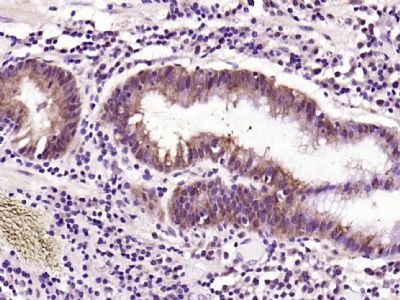 , ,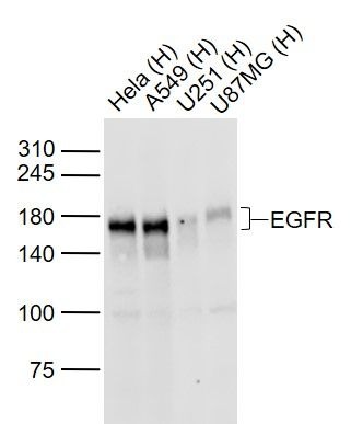 , ,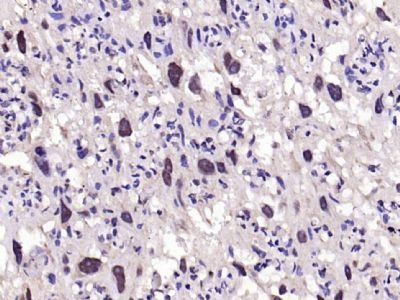 , ,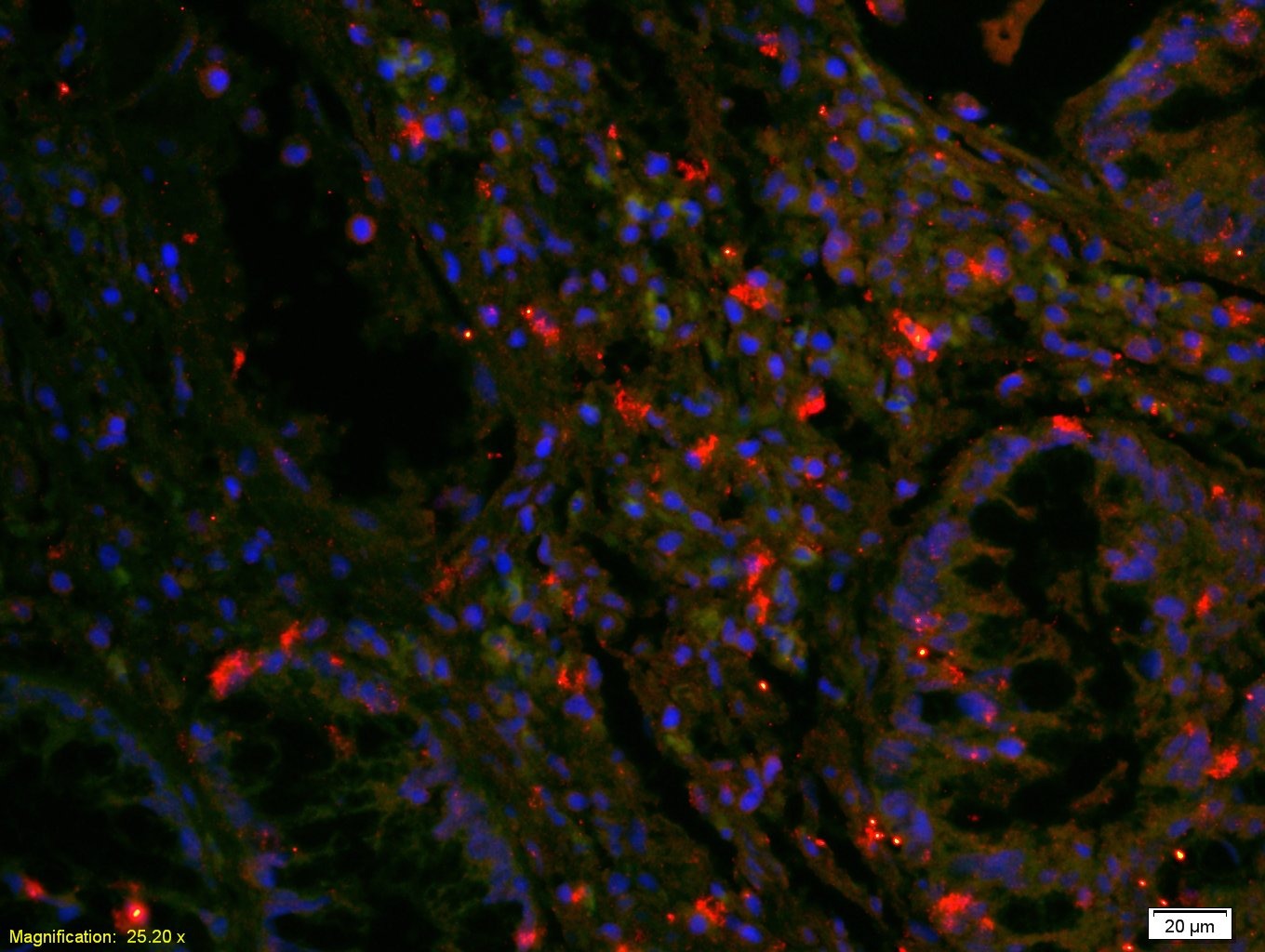 , ,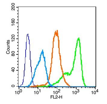 , ,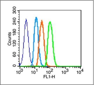 , ,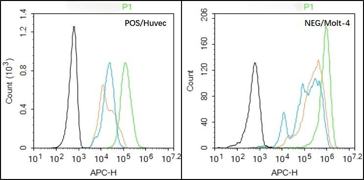 |
| Application | |
| Recommended Dose | WB: 1:500-2000; IHC-P: 1:100-500; IHC-Fr: 1:100-500; IF: 1:100-500; FCM: 1μg/Test |
| Antibody Type | Polyclonal |
| Host Species | Rabbit |
| Subcellular Localization | Cell membrane; Single-pass type I membrane protein. Endoplasmic reticulum membrane; Single-pass type I membrane protein. Golgi apparatus membrane; Single-pass type I membrane protein. Nucleus membrane; Single-pass type I membrane protein. Endosome. Endosome membrane. Note=In response to EGF, translocated from the cell membrane to the nucleus via Golgi and ER. Endocytosed upon activation by ligand. Isoform 2: Secreted. |
| Tissue Specificity | Ubiquitously expressed. Isoform 2 is also expressed in ovarian cancers. |
| Construction | Polyclonal Antibody |
| Purification | Protein A purified |
| Appearance | Liquid |
| Formulation | 0.01M TBS (pH7.4) with 1% BSA, 0.02% Proclin300 and 50% Glycerol. |
| Concentration | 1 mg/mL |
| Research Background | The protein encoded by this gene is a transmembrane glycoprotein that is a member of the protein kinase superfamily. This protein is a receptor for members of the epidermal growth factor family. EGFR is a cell surface protein that binds to epidermal growth factor. Binding of the protein to a ligand induces receptor dimerization and tyrosine autophosphorylation and leads to cell proliferation. Mutations in this gene are associated with lung cancer. Multiple alternatively spliced transcript variants that encode different protein isoforms have been found for this gene. [provided by RefSeq, Jul 2010] |
| Immunogen | KLH conjugated synthetic peptide: human EGFR |
| Antigen Species | Human |
| Gene Name | EGFR |
| Gene ID | |
| Protein Name | Epidermal growth factor receptor |
| Uniprot ID | |
| Biology Area | Receptor Tyrosine Kinases,EGF,egfr,SARS Coronavirus,Cell differentiation,Receptors,Receptor tyrosine kinases,Growth factor receptors,EGF |
| Function | Receptor tyrosine kinase binding ligands of the EGF family and activating several signaling cascades to convert extracellular cues into appropriate cellular responses. Known ligands include EGF, TGFA/TGF-alpha, amphiregulin, epigen/EPGN, BTC/betacellulin, epiregulin/EREG and HBEGF/heparin-binding EGF. Ligand binding triggers receptor homo- and/or heterodimerization and autophosphorylation on key cytoplasmic residues. The phosphorylated receptor recruits adapter proteins like GRB2 which in turn activates complex downstream signaling cascades. Activates at least 4 major downstream signaling cascades including the RAS-RAF-MEK-ERK, PI3 kinase-AKT, PLCgamma-PKC and STATs modules. May also activate the NF-kappa-B signaling cascade. Also directly phosphorylates other proteins like RGS16, activating its GTPase activity and probably coupling the EGF receptor signaling to the G protein-coupled receptor signaling. Also phosphorylates MUC1 and increases its interaction with SRC and CTNNB1/beta-catenin. Isoform 2 may act as an antagonist of EGF action. |
| Molecular Weight | Theoretical: 130 kDa. Actual: 170 kDa. |
| Stability & Storage | Store at -20°C or -80°C for 12 months. Avoid repeated freeze-thaw cycles. |
| Transport | Shipping with blue ice. |
| Size | Quantity | Unit Price | Amount | Operation |
|---|
 Hello! How can I help you today?
 Copyright © 2015-2026 TargetMol Chemicals Inc. All Rights Reserved.
