
Shopping Cart
Remove All Your shopping cart is currently empty

Synonyms: FSP27, FLJ20871, Fat-specific protein FSP27 homolog, Fat specific protein 27, CIDEC, CIDE3, CIDE C, CIDE 3, Cell death-inducing DFFA-like effector protein C, Cell Death Inducing DFFA Like Effector C, Cell death activator CIDE-3, Cell Death Activator
Anti-CIDEC Polyclonal Antibody
| Pack Size | Price | USA Stock | Global Stock | Quantity |
|---|---|---|---|---|
| 50 µL | $221 | 7-10 days | 7-10 days | |
| 100 µL | $373 | 7-10 days | 7-10 days | |
| 200 µL | $528 | 7-10 days | 7-10 days |
| Description | Anti-CIDEC Polyclonal Antibody is a Rabbit antibody targeting CIDEC. Anti-CIDEC Polyclonal Antibody can be used in FCM, ICC/IF, IF, IHC-Fr, IHC-P, WB. |
| Synonyms | FSP27, FLJ20871, Fat-specific protein FSP27 homolog, Fat specific protein 27, CIDEC, CIDE3, CIDE C, CIDE 3, Cell death-inducing DFFA-like effector protein C, Cell Death Inducing DFFA Like Effector C, Cell death activator CIDE-3, Cell Death Activator |
| Ig Type | IgG |
| Reactivity | Human,Mouse,Rat (predicted:Pig) |
| Verified Activity | 1. Paraformaldehyde-fixed, paraffin embedded (Rat brain); Antigen retrieval by boiling in sodium citrate buffer (pH6.0) for 15 min; Block endogenous peroxidase by 3% hydrogen peroxide for 20 min; Blocking buffer (normal goat serum) at 37°C for 30 min; Antibody incubation with (CIDEC) Polyclonal Antibody, Unconjugated (TMAB-00432) at 1:400 overnight at 4°C, followed by operating according to SP Kit (Rabbit) instructionsand DAB staining. 2. Tissue/cell: rat heart tissue; 4% Paraformaldehyde-fixed and paraffin-embedded; Antigen retrieval: citrate buffer (0.01M, pH6.0), Boiling bathing for 15 min; Block endogenous peroxidase by 3% Hydrogen peroxide for 30 min; Blocking buffer (normal goat serum) at 37°C for 20 min; Incubation: Anti-CIDEC Polyclonal Antibody, Unconjugated (TMAB-00432) 1:200, overnight at 4°C, followed by conjugation to the secondary antibody and DAb staining. 3. Tissue/cell: mouse stomach wall; 4% Paraformaldehyde-fixed and paraffin-embedded; Antigen retrieval: citrate buffer (0.01M, pH6.0), Boiling bathing for 15 min; Block endogenous peroxidase by 3% Hydrogen peroxide for 30 min; Blocking buffer (normal goat serum) at 37°C for 20 min; Incubation: Anti-CIDEC Polyclonal Antibody, Unconjugated (TMAB-00432) 1:200, overnight at 4°C, followed by conjugation to the secondary antibody and DAb staining. 4. Blank control: Mouse spleen. Primary Antibody (green line): Rabbit Anti-CIDEC antibody (TMAB-00432) Dilution: 2 μg/10^6 cells; Isotype Control Antibody (orange line): Rabbit IgG. Secondary Antibody: Goat anti-rabbit IgG-AF647 Dilution: 1 μg/test. Protocol The cells were fixed with 4% PFA (10 min at room temperature) and then permeabilized with 90% ice-cold methanol for 20 min at-20°C. The cells were then incubated in 5% BSA to block non-specific protein-protein interactions for 30 min at room temperature. Cells stained with Primary Antibody for 30 min at room temperature. The secondary antibody used for 40 min at room temperature. 5. Paraformaldehyde-fixed, paraffin embedded (rat breast); Antigen retrieval by boiling in sodium citrate buffer (pH6.0) for 15 min; Block endogenous peroxidase by 3% hydrogen peroxide for 20 min; Blocking buffer (normal goat serum) at 37°C for 30 min; Antibody incubation with (CIDEC) Polyclonal Antibody, Unconjugated (TMAB-00432) at 1:200 overnight at 4°C, followed by operating according to SP Kit (Rabbit) instructionsand DAB staining. 6. Paraformaldehyde-fixed, paraffin embedded (Mouse colon); Antigen retrieval by boiling in sodium citrate buffer (pH6.0) for 15 min; Block endogenous peroxidase by 3% hydrogen peroxide for 20 min; Blocking buffer (normal goat serum) at 37°C for 30 min; Antibody incubation with (CIDEC) Polyclonal Antibody, Unconjugated (TMAB-00432) at 1:200 overnight at 4°C, followed by operating according to SP Kit (Rabbit) instructionsand DAB staining. 7. Paraformaldehyde-fixed, paraffin embedded (rat colon); Antigen retrieval by boiling in sodium citrate buffer (pH6.0) for 15 min; Block endogenous peroxidase by 3% hydrogen peroxide for 20 min; Blocking buffer (normal goat serum) at 37°C for 30 min; Antibody incubation with (CIDEC) Polyclonal Antibody, Unconjugated (TMAB-00432) at 1:200 overnight at 4°C, followed by operating according to SP Kit (Rabbit) instructionsand DAB staining. 8. Paraformaldehyde-fixed, paraffin embedded (human gastric); Antigen retrieval by boiling in sodium citrate buffer (pH6.0) for 15 min; Block endogenous peroxidase by 3% hydrogen peroxide for 20 min; Blocking buffer (normal goat serum) at 37°C for 30 min; Antibody incubation with (CIDEC) Polyclonal Antibody, Unconjugated (TMAB-00432) at 1:200 overnight at 4°C, followed by operating according to SP Kit (Rabbit) instructionsand DAB staining. 9. Paraformaldehyde-fixed, paraffin embedded (rat brain); Antigen retrieval by boiling in sodium citrate buffer (pH6.0) for 15 min; Block endogenous peroxidase by 3% hydrogen peroxide for 20 min; Blocking buffer (normal goat serum) at 37°C for 30 min; Antibody incubation with (CIDEC) Polyclonal Antibody, Unconjugated (TMAB-00432) at 1:200 overnight at 4°C, followed by operating according to SP Kit (Rabbit) instructionsand DAB staining. 10. Paraformaldehyde-fixed, paraffin embedded (mouse brain); Antigen retrieval by boiling in sodium citrate buffer (pH6.0) for 15 min; Block endogenous peroxidase by 3% hydrogen peroxide for 20 min; Blocking buffer (normal goat serum) at 37°C for 30 min; Antibody incubation with (CIDEC) Polyclonal Antibody, Unconjugated (TMAB-00432) at 1:200 overnight at 4°C, followed by operating according to SP Kit (Rabbit) instructionsand DAB staining. 11. Paraformaldehyde-fixed, paraffin embedded (Human colon carcinoma); Antigen retrieval by boiling in sodium citrate buffer (pH6.0) for 15 min; Block endogenous peroxidase by 3% hydrogen peroxide for 20 min; Blocking buffer (normal goat serum) at 37°C for 30 min; Antibody incubation with (CIDEC) Polyclonal Antibody, Unconjugated (TMAB-00432) at 1:400 overnight at 4°C, followed by operating according to SP Kit (Rabbit) instructionsand DAB staining. 12. Blank control: A431. Primary Antibody (green line): Rabbit Anti-CIDEC antibody (TMAB-00432) Dilution: 1 μg/Test; Secondary Antibody: Goat anti-rabbit IgG-FITC Dilution: 0.5 μg/Test. Protocol The cells were fixed with 4% PFA (10 min at room temperature) and then permeabilized with 0.1% PBST for 20 min at room temperature. The cells were then incubated in 5% BSA to block non-specific protein-protein interactions for 30 min at room temperature. Cells stained with Primary Antibody for 30 min at room temperature. The secondary antibody used for 40 min at room temperature. 13. Sample: Lane 1: Adipose (Mouse) Lysate at 40 μg Lane 2: Breast (Mouse) Lysate at 40 μg Lane 3: Adipose (Rat) Lysate at 40 μg Primary: Anti-CIDEC (TMAB-00432) at 1/1000 dilution Secondary: IRDye800CW Goat Anti-Rabbit IgG at 1/20000 dilution Predicted band size: 27-30 kDa Observed band size: 27 kDa 14. Sample: Epididymis (Mouse) Lysate at 40 μg Primary: Anti-CIDEC (TMAB-00432) at 1/500 dilution Secondary: IRDye800CW Goat Anti-Rabbit IgG at 1/20000 dilution Predicted band size: 27 kDa Observed band size: 27 kDa 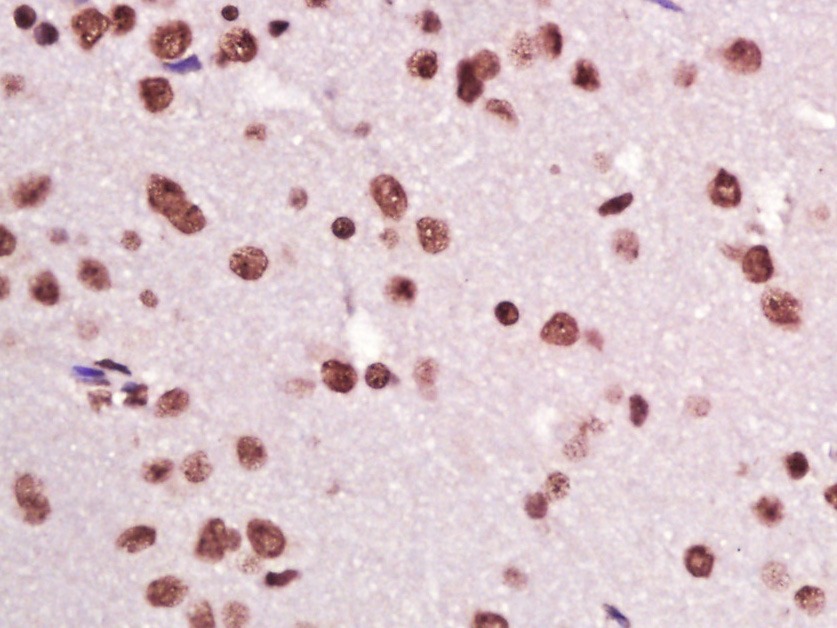 , ,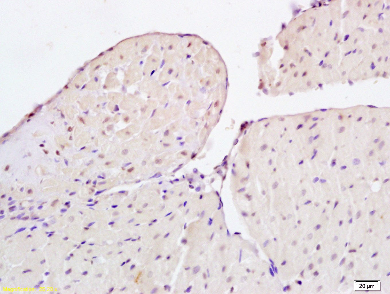 , ,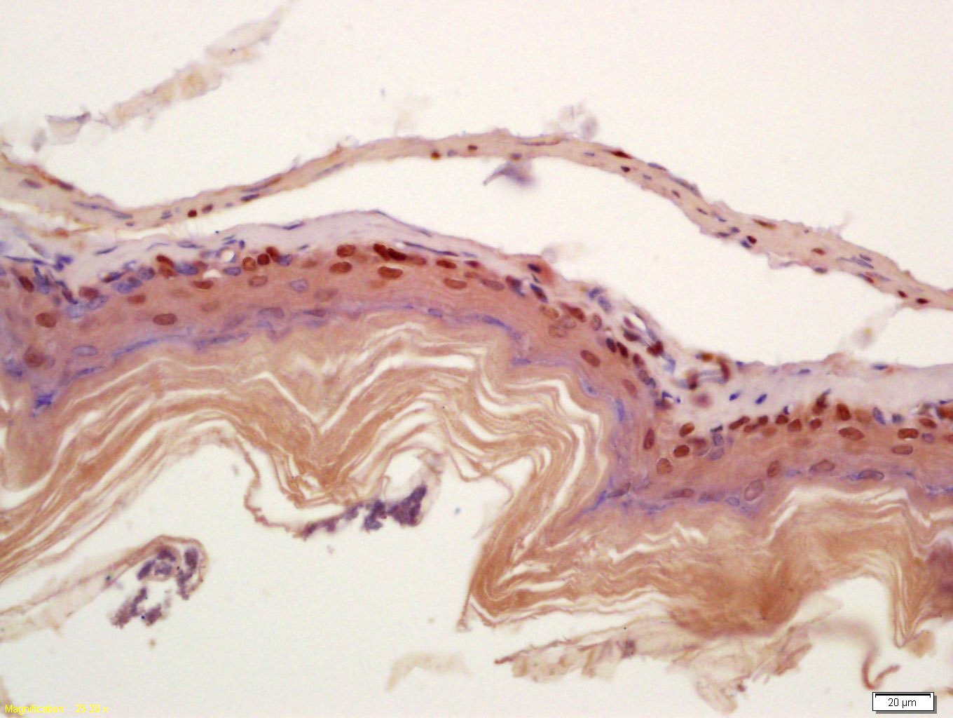 , ,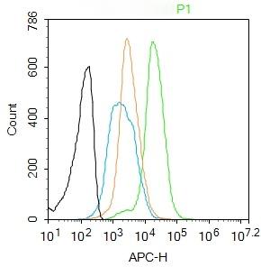 , ,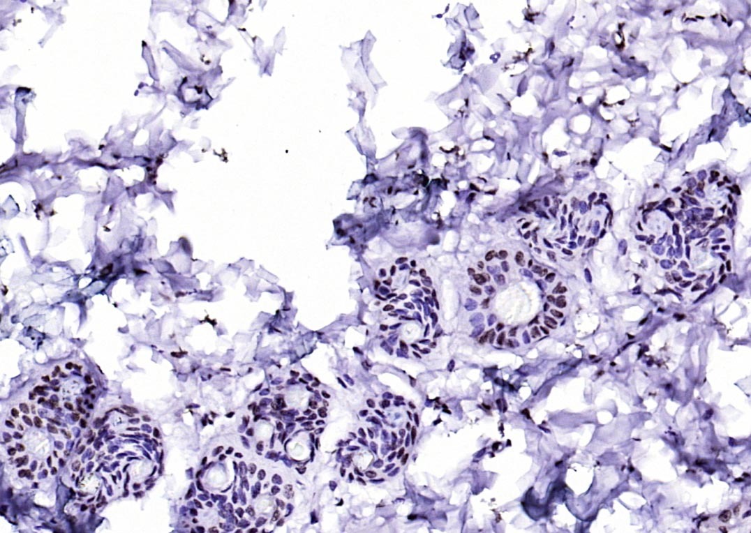 , ,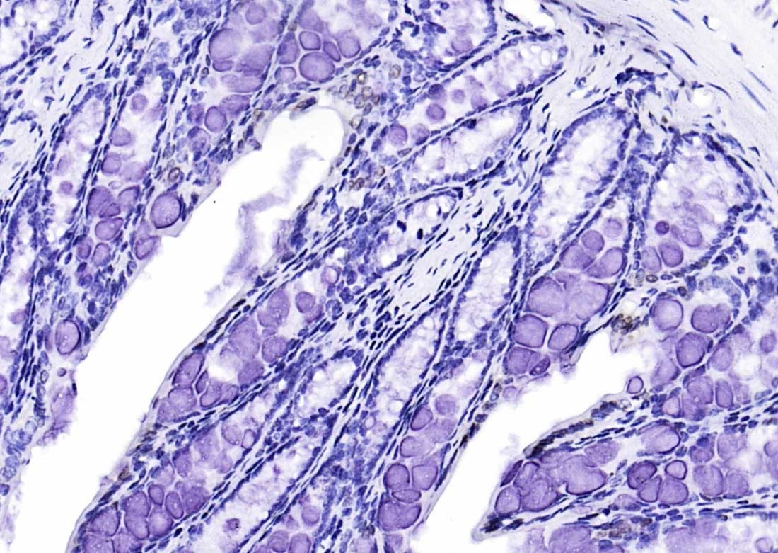 , ,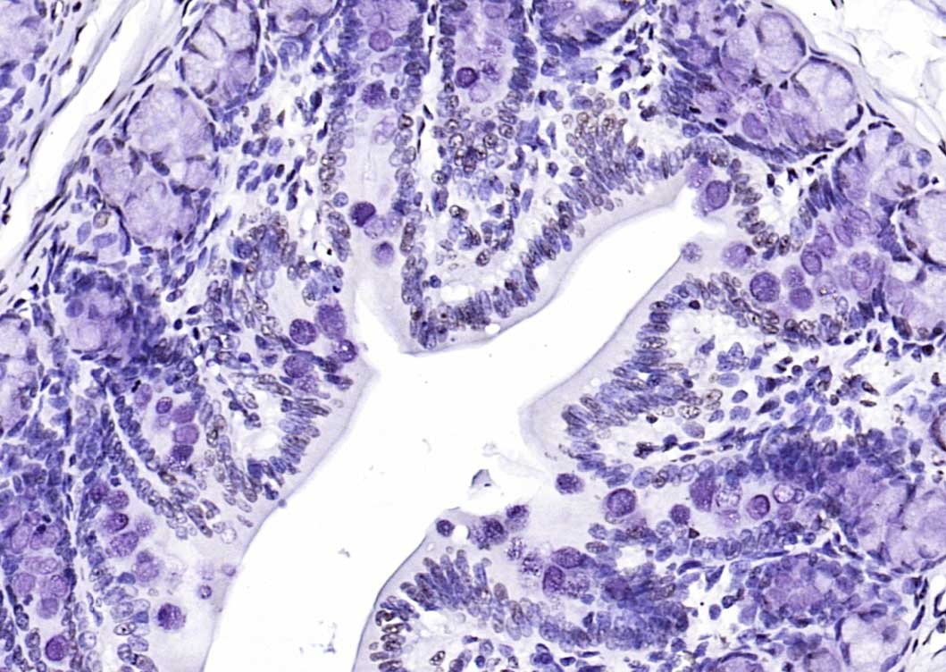 , ,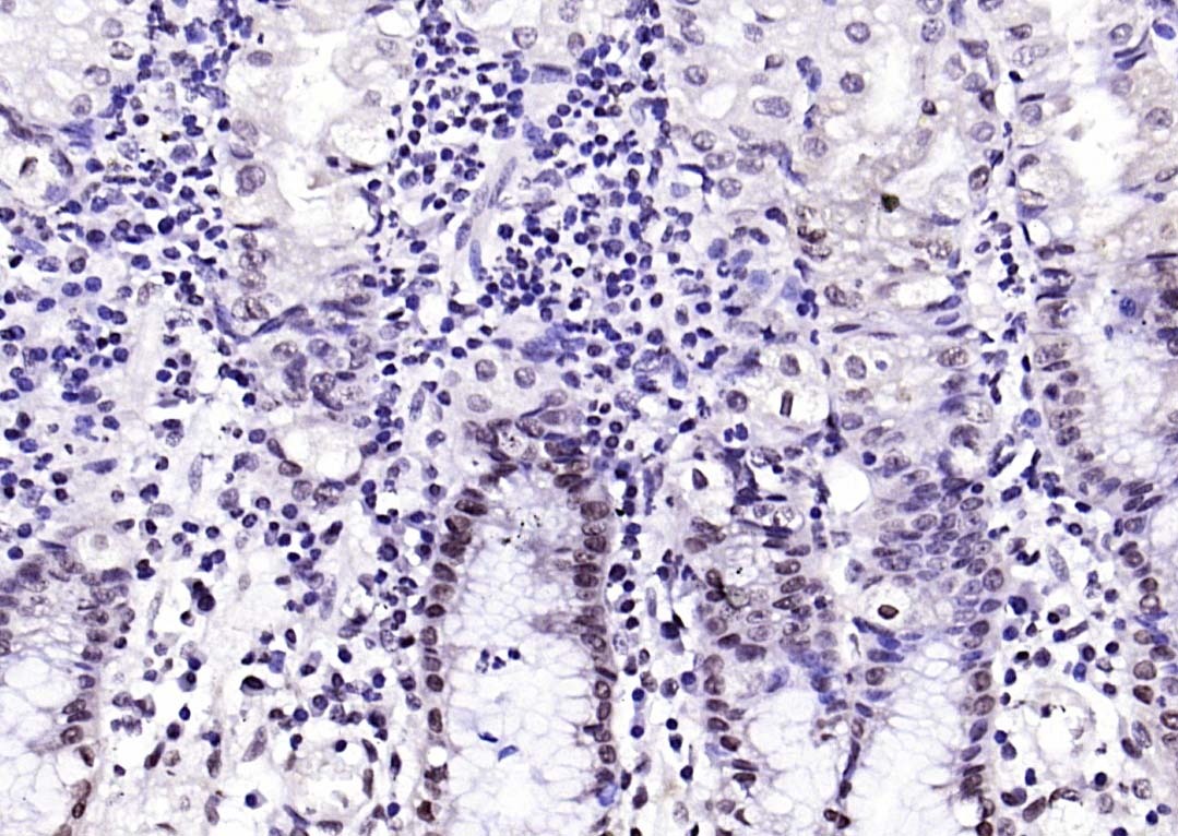 , ,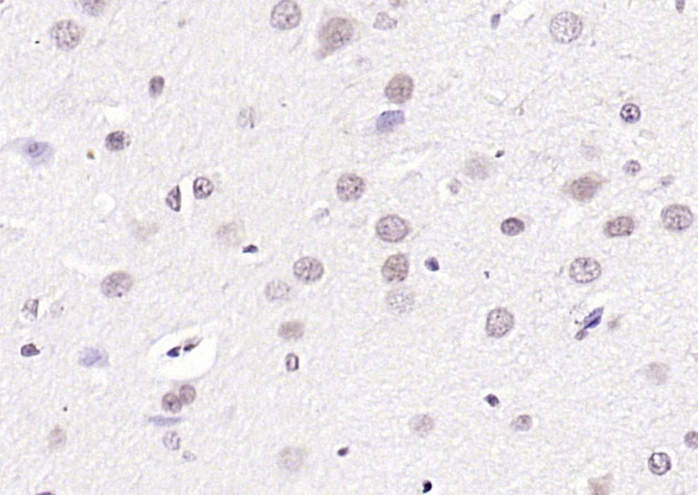 , ,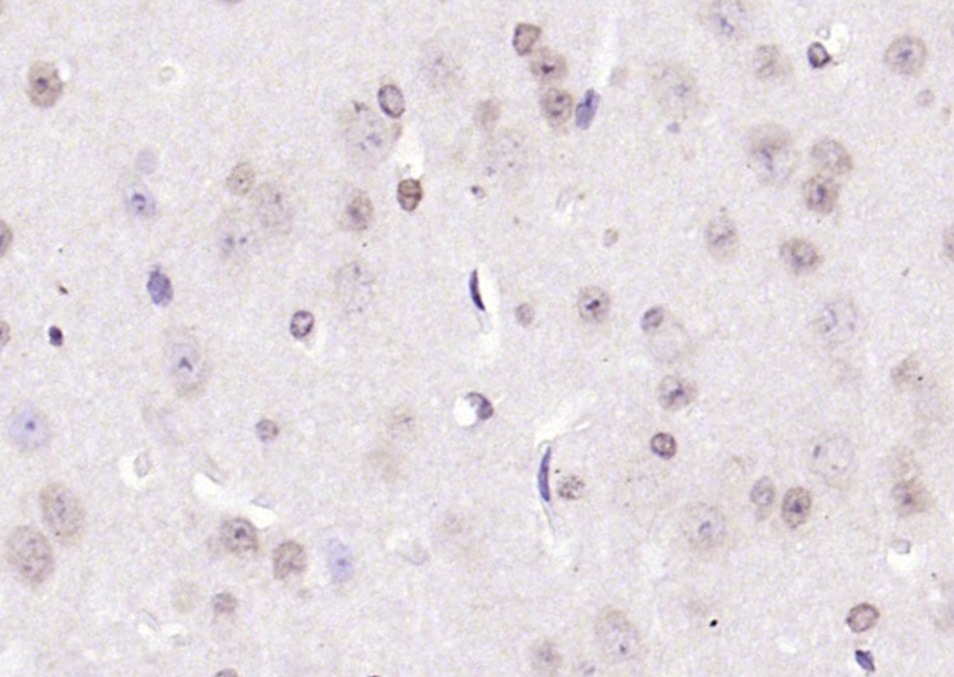 , ,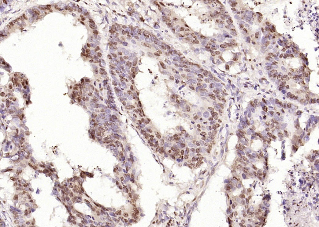 , ,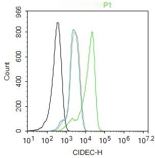 , ,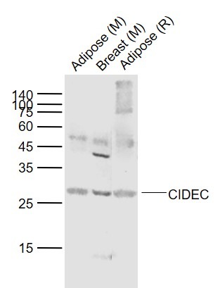 , ,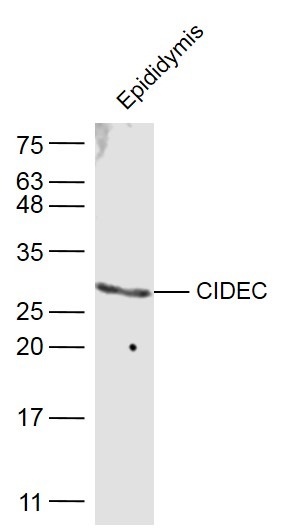 |
| Application | |
| Recommended Dose | FCM=1 μg/Test; ICC/IF=1:50-200; IF=1:100-500; IHC-Fr=1:100-500; IHC-P=1:100-500; WB=1:500-2000 |
| Antibody Type | Polyclonal |
| Host Species | Rabbit |
| Subcellular Localization | Nucleus (By similarity). Endoplasmicreticulum (By similarity). Lipid droplet. Note=Diffuses quickly onlipid droplet surface, but becomes trapped and clustered at lipiddroplet contact sites, thereby enabling its rapid enrichment atlipid droplet contact sites. |
| Tissue Specificity | Expressed mainly in adipose tissue, smallintestine, heart, colon and stomach and, at lower levels, in brain,kidney and liver. |
| Construction | Polyclonal Antibody |
| Purification | Protein A purified |
| Appearance | Liquid |
| Formulation | 0.01M TBS (pH7.4) with 1% BSA, 0.02% Proclin300 and 50% Glycerol. |
| Concentration | 1 mg/mL |
| Research Background | This gene encodes a member of the cell death-inducing DNA fragmentation factor-like effector family. Members of this family play important roles in apoptosis. The encoded protein promotes lipid droplet formation in adipocytes and may mediate adipocyte apoptosis. This gene is regulated by insulin and its expression is positively correlated with insulin sensitivity. Mutations in this gene may contribute to insulin resistant diabetes. A pseudogene of this gene is located on the short arm of chromosome 3. Alternatively spliced transcript variants that encode different isoforms have been observed for this gene. [provided by RefSeq, Dec 2010]. Tissue specificity: Expressed mainly in small intestine, heart, colon and stomach and, at lower levels, in brain, kidney and liver. |
| Immunogen | KLH conjugated synthetic peptide: human CIDEC |
| Antigen Species | Human |
| Gene Name | CIDEC |
| Gene ID | |
| Protein Name | Cell death activator CIDE-3 |
| Uniprot ID | |
| Function | May act as a CEBPB coactivator in white adipose tissueto control the expression of a subset of CEBPB downstream targetgenes, including SOCS1, SOCS3, TGFB1, TGFBR1, ID2 and XDH (Bysimilarity). Binds to lipid droplets and regulates theirenlargement, thereby restricting lipolysis and favoring storage. Atfocal contact sites between lipid droplets, promotes directionalnet neutral lipid transfer from the smaller to larger lipiddroplets. The transfer direction may be driven by the internalpressure difference between the contacting lipid droplet pair. Whenoverexpressed in preadipocytes, induces apoptosis or increases cellsusceptibility to apoptosis induced by serum deprivation or TGFBtreatment. As mature adipocytes, that express high CIDEC levels,are quite resistant to apoptotic stimuli, the physiologicalsignificance of its role in apoptosis is unclear. |
| Molecular Weight | Theoretical: 27 kDa. Actual: 27 kDa. |
| Stability & Storage | Store at -20°C or -80°C for 12 months. Avoid repeated freeze-thaw cycles. |
| Transport | Shipping with blue ice. |
| Size | Quantity | Unit Price | Amount | Operation |
|---|
 Hello! How can I help you today?
 Copyright © 2015-2026 TargetMol Chemicals Inc. All Rights Reserved.
