
Shopping Cart
Remove All Your shopping cart is currently empty

Synonyms: P90, IP90, CNX, calnexin
Anti-Calnexin Antibody
(2Q119)
| Pack Size | Price | USA Stock | Global Stock | Quantity |
|---|---|---|---|---|
| 50 µL | $298 | 7-10 days | 7-10 days | |
| 100 µL | $496 | 7-10 days | 7-10 days |
| Description | Anti-Calnexin Antibody (2Q119) is a Rabbit antibody targeting Calnexin. Anti-Calnexin Antibody (2Q119) can be used in FCM,ICC/IF,IHC,WB. |
| Synonyms | P90, IP90, CNX, calnexin |
| Ig Type | IgG |
| Clone | 2Q119 |
| Reactivity | Human,Rat |
| Verified Activity | 1. Western blot analysis of Calnexin on Hela cells lysates using anti-Calnexin antibody at 1/1,000 dilution. 2. Immunohistochemical analysis of paraffin-embedded human pancreas tissue using anti-Calnexin antibody. Counter stained with hematoxylin. 3. Immunohistochemical analysis of paraffin-embedded rat heart tissue using anti-Calnexin antibody. Counter stained with hematoxylin. 4. Immunohistochemical analysis of paraffin-embedded rat pancreas tissue using anti-Calnexin antibody. Counter stained with hematoxylin. 5. Immunohistochemical analysis of paraffin-embedded human liver cancer tissue using anti-Calnexin antibody. Counter stained with hematoxylin. 6. Immunohistochemical analysis of paraffin-embedded rat kidney tissue using anti-Calnexin antibody. Counter stained with hematoxylin. 7. Immunohistochemical analysis of paraffin-embedded human kidney tissue using anti-Calnexin antibody. Counter stained with hematoxylin. 8. ICC staining Calnexin in Hela cells (green). The nuclear counter stain is DAPI (blue). Cells were fixed in paraformaldehyde, permeabilised with 0.25% Triton X100/PBS. 9. ICC staining Calnexin in HepG2 cells (green). The nuclear counter stain is DAPI (blue). Cells were fixed in paraformaldehyde, permeabilised with 0.25% Triton X100/PBS. 10. ICC staining Calnexin in PANC-1 cells (green). The nuclear counter stain is DAPI (blue). Cells were fixed in paraformaldehyde, permeabilised with 0.25% Triton X100/PBS. 11. Flow cytometric analysis of Hela cells with Calnexin antibody at 1/50 dilution (red) compared with an unlabelled control (cells without incubation with primary antibody; black). Alexa Fluor 488-conjugated goat anti rabbit IgG was used as the secondary antibody. 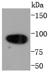 , ,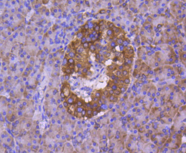 , ,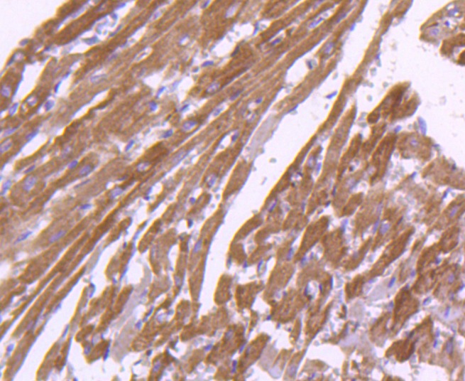 , ,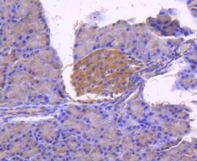 , ,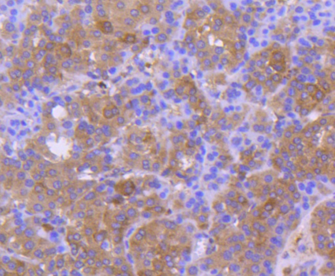 , ,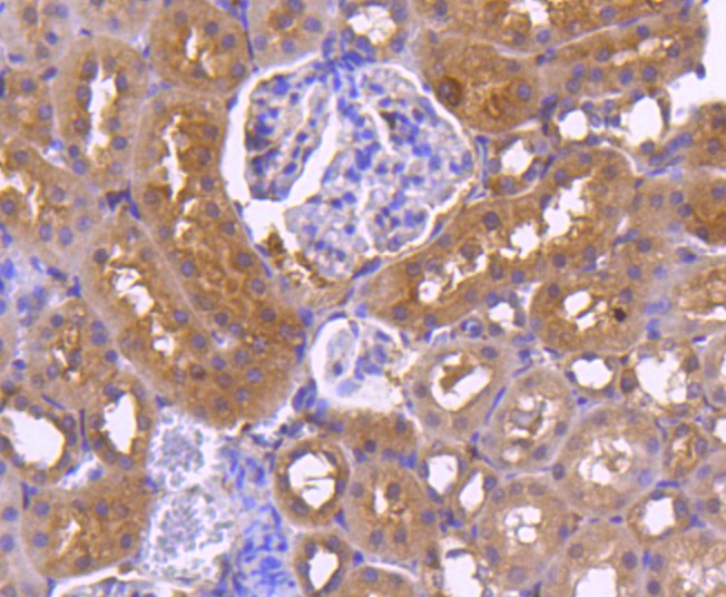 , ,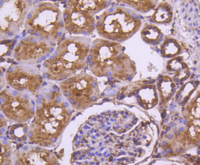 , ,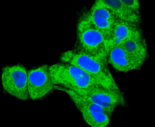 , ,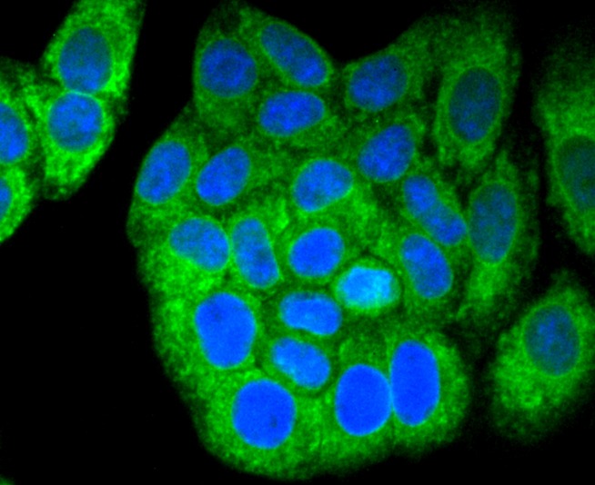 , ,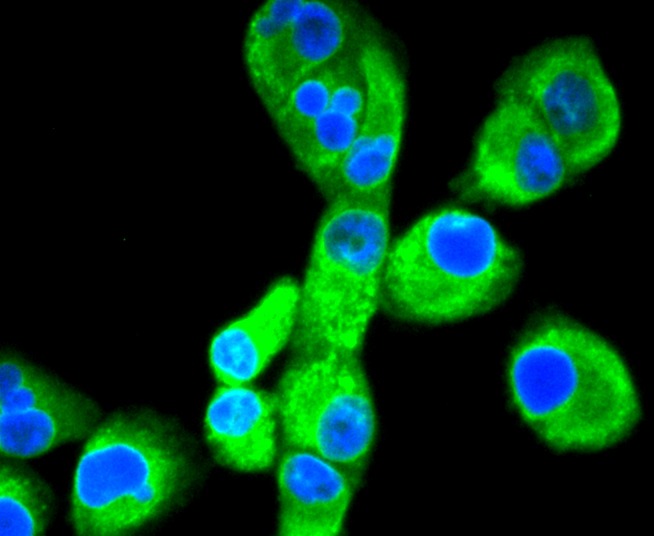 , ,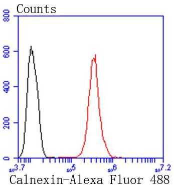 |
| Application | |
| Recommended Dose | WB: 1:1000-5000; IHC: 1:50-200; ICC/IF: 1:100-500; FCM: 1:50-100 |
| Antibody Type | Monoclonal |
| Host Species | Rabbit |
| Construction | Recombinant Antibody |
| Purification | ProA affinity purified |
| Appearance | Liquid |
| Formulation | 1*TBS (pH7.4), 1%BSA, 40%Glycerol. Preservative: 0.05% Sodium Azide. |
| Research Background | Calnexin and Calregulin (also called calreticulin) are calcium-binding proteins that are localized to the endoplasmic reticulum, Calnexin to the membrane and Calregulin to the lumen. Calnexin is a type I membrane protein that interacts with newly synthesized glycoproteins in the endoplasmic reticulum. It may play a role in assisting with protein assembly and in retaining unassembled protein subunits in the endoplasmic reticulum. Calregulin has both low- and high-affinity calcium-binding sites. Neither Calnexin nor Calregulin contains the calcium-binding ??E-F hand?? motif found in calmodulins. Calnexin and Calregulin are important for the maturation of glycoproteins in the endoplasmic reticulum and appear to bind many of the same proteins. |
| Conjucates | Unconjugated |
| Immunogen | Recombinant Protein |
| Uniprot ID |
| Molecular Weight | Theoretical: 90 kDa. |
| Stability & Storage | Store at -20°C or -80°C for 12 months. Avoid repeated freeze-thaw cycles. |
| Transport | Shipping with blue ice. |
| Size | Quantity | Unit Price | Amount | Operation |
|---|
 Hello! How can I help you today?
 Copyright © 2015-2026 TargetMol Chemicals Inc. All Rights Reserved.
