
Shopping Cart
Remove All Your shopping cart is currently empty

Synonyms: Pro apoptotic protein BAK, NBak, MGC3887, MGC117255, Cell death inhibitor 1, CDN1, CDN 1, BCL2L7, BCL2-antagonist/killer 1, Bcl2 like 7 Protein, Bcl2 homologous antagonist killer, Bcl 2 like 7 protein, Bcl 2 homologous antagonist/killer, BAK1, Bak NT, BAK like, BAK 1, BAK, Apoptosis Regulator Bak
Anti-BAK1 Polyclonal Antibody
| Pack Size | Price | USA Stock | Global Stock | Quantity |
|---|---|---|---|---|
| 50 µL | $222 | 7-10 days | 7-10 days | |
| 100 µL | $374 | 7-10 days | 7-10 days | |
| 200 µL | $528 | 7-10 days | 7-10 days |
| Description | Anti-BAK1 Polyclonal Antibody is a Rabbit antibody targeting BAK1. Anti-BAK1 Polyclonal Antibody can be used in FCM,IF,IHC-Fr,IHC-P,WB. |
| Synonyms | Pro apoptotic protein BAK, NBak, MGC3887, MGC117255, Cell death inhibitor 1, CDN1, CDN 1, BCL2L7, BCL2-antagonist/killer 1, Bcl2 like 7 Protein, Bcl2 homologous antagonist killer, Bcl 2 like 7 protein, Bcl 2 homologous antagonist/killer, BAK1, Bak NT, BAK like, BAK 1, BAK, Apoptosis Regulator Bak |
| Ig Type | IgG |
| Reactivity | Human,Mouse (predicted:Rat) |
| Verified Activity | 1. Tissue/cell: human colon carcinoma; 4% Paraformaldehyde-fixed and paraffin-embedded; Antigen retrieval: citrate buffer (0.01M, pH6.0), Boiling bathing for 15 min; Block endogenous peroxidase by 3% Hydrogen peroxide for 30 min; Blocking buffer (normal goat serum) at 37°C for 20 min; Incubation: Anti-Bak Polyclonal Antibody, Unconjugated (TMAB-00183) 1:200, overnight at 4°C, followed by conjugation to the secondary antibody and DAb staining. 2. Sample: Lung (Mouse) Lysate at 30 μg Primary: Anti-Bak (TMAB-00183) at 1:300 dilution; Secondary: HRP conjugated Goat-Anti-Rabbit Igg (bse-0295G) at 1: 5000 dilution; Predicted band size: 23 kDa Observed band size: 23 kDa 3. Tissue/cell: human lung carcinoma; 4% Paraformaldehyde-fixed and paraffin-embedded; Antigen retrieval: citrate buffer (0.01M, pH6.0), Boiling bathing for 15 min; Block endogenous peroxidase by 3% Hydrogen peroxide for 30 min; Blocking buffer (normal goat serum) at 37°C for 20 min; Incubation: Anti-Bak Polyclonal Antibody, Unconjugated (TMAB-00183) 1:500, overnight at 4°C, followed by conjugation to the secondary antibody and DAb staining. 4. Blank control: Hela (blue). Primary Antibody: Rabbit Anti-Bak antibody, Dilution: 1 μg in 100 μL 1X PBS containing 0.5% BSA; Isotype Control Antibody: Rabbit Igg (orange),used under the same conditions); Secondary Antibody: Goat anti-rabbit IgG-Pe (white blue), Dilution: 1:200 in 1 X PBS containing 0.5% BSA. Protocol The cells were fixed with 2% paraformaldehyde (10 min), then permeabilized with 90% ice-cold methanol for 30 min on ice. Antibody (TMAB-00183, 1 μg/1x10^6 cells) were incubated for 30 min on the ice, followed by 1 X PBS containing 0.5% BSA + 10% goat serum (15 min) to block non-specific protein-protein interactions. Then the Goat Anti-rabbit IgG/PE antibody was added into the blocking buffer mentioned above to react with the primary antibody of TMAB-00183 at 1/200 dilution for 30 min on ice. 5. Blank control: THP-1. Primary Antibody (green line): Rabbit Anti-Bak antibody (TMAB-00183) Dilution: 2 μg/10^6 cells; Isotype Control Antibody (orange line): Rabbit IgG. Secondary Antibody: Goat anti-rabbit IgG-FITC Dilution: 1 μg/test. Protocol The cells were fixed with 4% PFA (10 min at room temperature) and then permeabilized with 0.1% PBST methanol for 20 min at room temperature. The cells were then incubated in 5% BSA to block non-specific protein-protein interactions for 30 min at room temperature. Cells stained with Primary Antibody for 30 min at room temperature. The secondary antibody used for 40 min at room temperature. 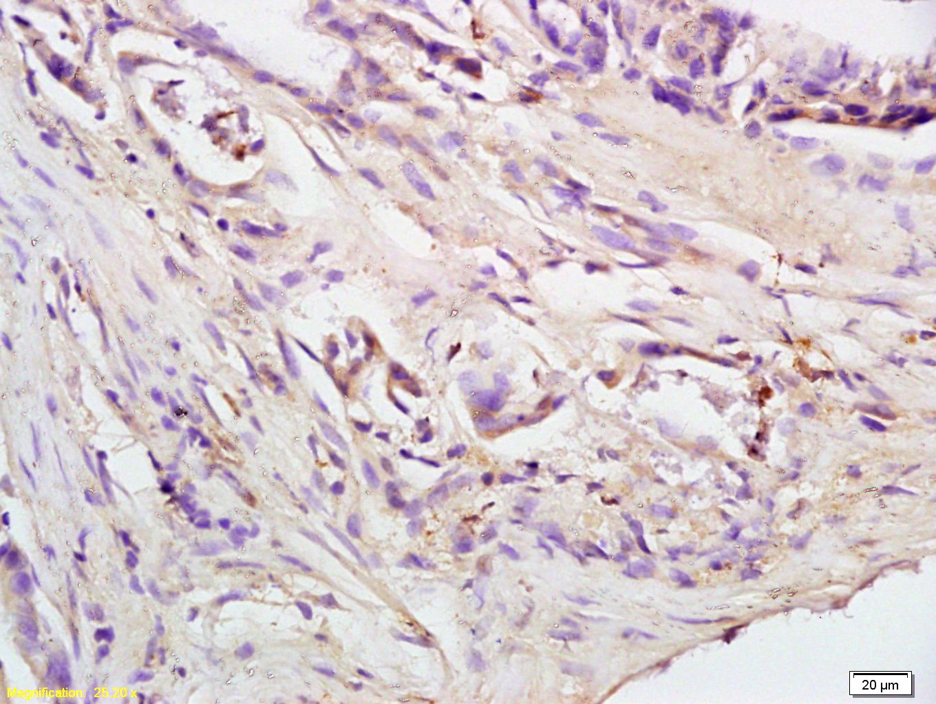 , ,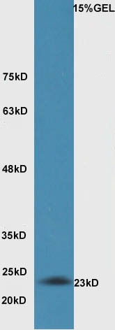 , ,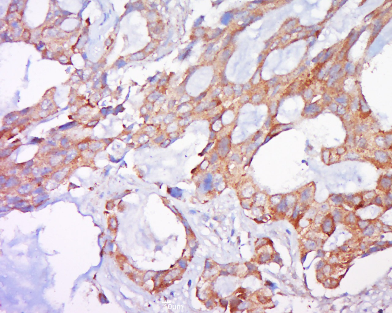 , ,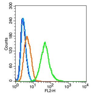 , ,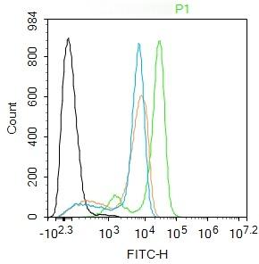 |
| Application | |
| Recommended Dose | WB: 1:500-2000; IHC-P: 1:100-500; IHC-Fr: 1:100-500; IF: 1:100-500; FCM: 1μg/Test |
| Antibody Type | Polyclonal |
| Host Species | Rabbit |
| Subcellular Localization | Mitochondrion membrane. |
| Tissue Specificity | Expressed in a wide variety of tissues. |
| Construction | Polyclonal Antibody |
| Purification | Protein A purified |
| Appearance | Liquid |
| Formulation | 0.01M TBS (pH7.4) with 1% BSA, 0.02% Proclin300 and 50% Glycerol. |
| Concentration | 1 mg/mL |
| Research Background | The protein encoded by this gene belongs to the BCL2 protein family. BCL2 family members form oligomers or heterodimers and act as anti- or pro-apoptotic regulators that are involved in a wide variety of cellular activities. This protein localizes to mitochondria, and functions to induce apoptosis. It interacts with and accelerates the opening of the mitochondrial voltage-dependent anion channel, which leads to a loss in membrane potential and the release of cytochrome c. This protein also interacts with the tumor suppressor P53 after exposure to cell stress. [provided by RefSeq, Jul 2008] |
| Immunogen | KLH conjugated synthetic peptide: mouse Bak |
| Antigen Species | Mouse |
| Gene Name | BAK1 |
| Gene ID | |
| Protein Name | Bcl-2 homologous antagonist/killer |
| Uniprot ID | |
| Biology Area | Bcl2 Family |
| Function | In the presence of an appropriate stimulus, accelerates programmed cell death by binding to, and antagonizing the anti-apoptotic action of BCL2 or its adenovirus homolog E1B 19k protein. Low micromolar levels of zinc ions inhibit the promotion of apoptosis. |
| Molecular Weight | Theoretical: 23 kDa. Actual: 23 kDa. |
| Stability & Storage | Store at -20°C or -80°C for 12 months. Avoid repeated freeze-thaw cycles. |
| Transport | Shipping with blue ice. |
| Size | Quantity | Unit Price | Amount | Operation |
|---|
 Hello! How can I help you today?
 Copyright © 2015-2026 TargetMol Chemicals Inc. All Rights Reserved.
